Download
1 / 43
520 likes | 1.13k Vues
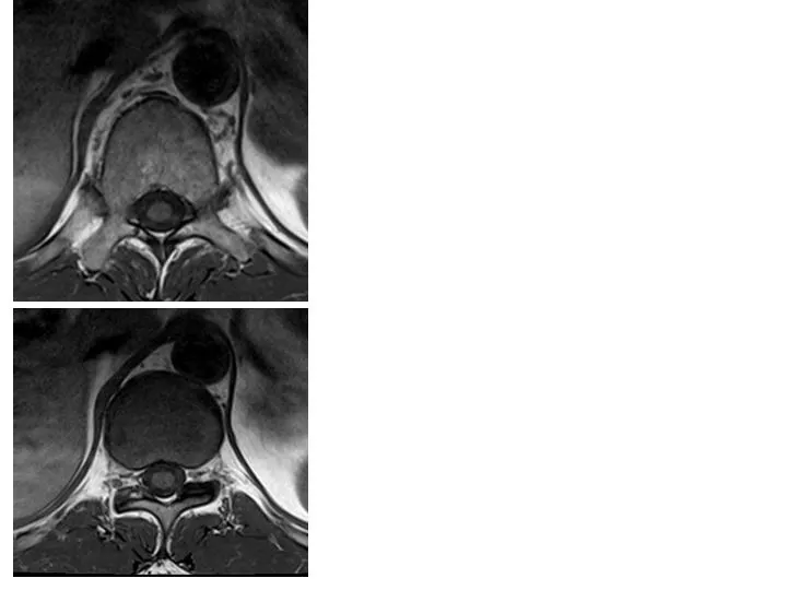
MRI of the Thoracic Spine: Axial T1 wtd.images. A : Yellow arrow: Thoracic cord Long white arrow: Cerebrospinal fluid space Orange arrow: Costotransverse joint Green arrow: Pedicle White arrowhead: Transverse process. T11 Body. Rib. A. B:

E N D
MRI of the Thoracic Spine: Axial T1 wtd.images. A : Yellow arrow: Thoracic cord Long white arrow: Cerebrospinal fluid space Orange arrow: Costotransverse joint Green arrow: Pedicle White arrowhead: Transverse process T11 Body Rib A B: White arrow: Neural foramina with exiting nerve root Yellow arrow: Facet joint Red arrow: Spinous process Green arrow : Lamina Orange arrow: Thoracic Aorta T11-T12 Disc B
Yellow arrow : Bright T2 wtd . CSF signal surrounds the cervical cord . Red arrow : Cervical cord. Blue arrow : Left C6 nerve root exits out of the spinal canal through C5-C6 neural foramina . White arrow : Vertebral artery . C5-C6 DISC Axial T2 wtd. MRI of the cervical spine at C5-C6 level
MRI of the Thoracic Spine : Sagittal T2 wtd. Image White arrow: Degenerative disc disease at T7-T8 level. Yellow arrow : Normal thoracic cord is seen surrounded by bright cerebrospinal fluid(C.S.F) signal on T2 wtd. pulse sequence, which is the sequence of choice to evaluate spinal cord pathology.
MRI cervical spine : Sagittal T2wtd. fat suppressed Image . C.S.F signal is bright ( yellow arrow ) on T2 wtd. Image ( dark on T1 wtd. Image ). Cerebro spinal fluid surrounds the spinal cord. Subcutaneous fat signal is suppressed and appears dark on fat suppressed image( green arrow ) whereas fat signal is bright on T1 wtd. Image . Red arrow points to cervical cord . C2 C5 T1
5 1 2 3 • MRI OF CERVICAL SPINE : Sagittal projection • T1 wtd. Image : CSF is dark ( yellow arrow ) • surrounding the thoracic cord ( red arrow ) • FAT is bright ( green arrow ) on T1 wtd. Image . • Anterior arch of C1 • Odontoid process of C2 • C2 vertebral body • C5-C6 disk space • Posterior arch of C1 • Cervical cord • C7 spinous process 6 C5 7 4 T1
4 5 6 7 8 9 1 2 3 11 12 10 13 24 14 Identify anatomical structures 1 - 24 18 19 17 20 21 16 22 15 23 Fig. 1.10 A midline Post-contrast Sagittal T1 Weighted MRI
3 Fig. 1.10 A midline Post-contrast Sagittal t1 Weighted MRI
Identify the brain structures in the following post-contrast axial MR images (Fig 1.1 to Fig 1.9) marked 1 through 45
Post Contrast sagittal T1 Weighted M.R.I. Section at the level of Foramen Magnum Fig. 1.1 Post Contrast Axial MR Image of the brain
Post Contrast sagittal T1 Wtd M.R.I. Section at the level of medulla Fig. 1.2 Post Contrast Axial MR Image of the brain
Post Contrast sagittal T1 Wtd M.R.I. Section at the level of Pons Fig. 1.3 Post Contrast Axial MR Image of the brain
Post Contrast sagittal T1 Wtd M.R.I. Section at the level of Mid Brain Fig. 1.4 Post Contrast Axial MR Image of the brain
Post Contrast sagittal T1 Wtd M.R.I. Section at the level of the III Ventricle Fig. 1.5 Post Contrast Axial MR Image of the brain
Post Contrast sagittal T1 Wtd M.R.I. Section at the level of Thalamus Fig. 1.6 Post Contrast Axial MR Image of the brain
Post Contrast sagittal T1 Wtd M.R.I. Section at the level of Corpus Callosum Fig. 1.7 Post Contrast Axial MR Image of the brain
Post Contrast sagittal T1 Wtd M.R.I. Section at the level of Body of Corpus Callosum Fig. 1.8 Post Contrast Axial MR Image of the brain
Post Contrast sagittal T1 Wtd M.R.I. Section above the Corpus Callosum Fig. 1.9 Post Contrast Axial MR Image of the brain
Identify the brain structures in the following post-contrast axial MR images (Fig 1.1 to Fig 1.9) marked 1 through 45
Fig. 1.11. Coronal Section of the Brain at the level of IV Ventricle Post Contrast Coronal T1 Weighted MRI
Fig. 1.12. Coronal Section of the Brain at the level of Pituitary gland Post Contrast Coronal T1 Weighted MRI
Fig. 1.13. Coronal Section of the Brain at the level of the orbits. Post Contrast Coronal T1 Weighted MRI.
4 5 6 7 8 9 1 2 3 11 12 10 13 24 14 1. Scalp fat 2. Bone 3. Inferior sagittal sinus 4. Corpus callosum 5. Internal cerebral vein 6. Vein of Galen 7. Superior sagittal sinus 8. Parietal lobe 9. Occipital lobe 10. Straight sinus 11. Vermis 12. IV ventricle 13. Cerebellar tonsil 14. Cervical cord 15. Medulla 16. Pons 17. Midbrain 18. Mass intermedia of thalamus 19. Anterior III ventricle 20. Optic chiasm 21. Pituitary gland 22. Sphenoid sinus 23. Nasopharynx 24. Frontal lobe 18 19 17 20 21 16 22 15 23 Fig. 1.10 A midline Post-contrast Sagittal T1 Weighted MRI
8 7 6 5 4 2 3 1 Identify anatomical structures 1 - 8 Fig. 1.11. Coronal Section of the Brain at the level of IV Ventricle Post Contrast Coronal T1 Weighted MRI
8 7 6 5 4 2 3 1 1. Cerebellar tonsil 2. Cerebellar hemisphere 3. IV ventricle 4. Superior vermis 5. Tentorium 6. Posterior temporal lobe 7. Choroid plexus within lateral ventricle 8. Posterior frontal lobe Fig. 1.11. Coronal Section of the Brain at the level of IV Ventricle Post Contrast Coronal T1 Weighted MRI
1 5 6 7 9 2 3 4 8 11 10 Identify anatomical structures 1 - 12 12 Fig. 1.12. Coronal Section of the Brain at the level of Pituitary gland Post Contrast Coronal T1 Weighted MRI
1 5 6 7 9 2 3 4 1. Frontal lobe 2. Corpus callosum 3. Frontal horn 4. Caudate nucleus 5. III ventricle 6. Optic nerve 7. Pituitary stalk 8. Pituitary gland 9. Internal carotid artery 10. Cavernous sinus 11. Sphenoid sinus 12. Nasopharynx 8 11 10 12 Fig. 1.12. Coronal Section of the Brain at the level of Pituitary gland Post Contrast Coronal T1 Weighted MRI
1 2 3 4 5 Identify anatomical structures 1 - 5 Fig. 1.13. Coronal Section of the Brain at the level of the orbits. Post Contrast Coronal T1 Weighted MRI.
1 2 3 4 5 1. Frontal lobe 2. Orbital Fat 3. Globe 4. Nasal Cavity 5. Maxillary Sinus Fig. 1.13. Coronal Section of the Brain at the level of the orbits. Post Contrast Coronal T1 Weighted MRI.
Identify the brain structures in the following post-contrast axial MR images (Fig 1.1 to Fig 1.9) marked 1 through 45
5 3 Post Contrast sagittal T1 Weighted M.R.I. Section at the level of Foramen Magnum 2 1 Answers 4 1. Cisterna Magna 2. Cervical Cord 3. Nasopharynx 4. Mandible 5. Maxillary Sinus Fig. 1.1 Post Contrast Axial MR Image of the brain
Post Contrast sagittal T1 Wtd M.R.I. Section at the level of medulla 6 Answers 6. Medulla 7. Sigmoid Sinus 7 Fig. 1.2 Post Contrast Axial MR Image of the brain
14 13 12 Post Contrast sagittal T1 Wtd M.R.I. Section at the level of Pons 11 17 10 Answers 16 15 9 13. Internal Carotid Artery 14. Cavernous Sinus 15. Middle Cerebellar Peduncle 16. Internal Auditory Canal 17. Temporal Lobe 8. Cerebellar Hemisphere 9. Vermis 10. IV Ventricle 11. Pons 12. Basilar Artery 8 Fig. 1.3 Post Contrast Axial MR Image of the brain
20 22 19 Post Contrast sagittal T1 Wtd M.R.I. Section at the level of Mid Brain 18 21 Answers 18. Aqueduct of Sylvius 19. Midbrain 20. Orbits 21. Posterior Cerebral Artery 22. Middle Cerebral Artery Fig. 1.4 Post Contrast Axial MR Image of the brain
27 25 24 Post Contrast sagittal T1 Wtd M.R.I. Section at the level of the III Ventricle Answers 23 23. Occipital Lobe 24. III Ventricle 25. Frontal Lobe 26. Temporal Lobe 27. Sylvian Fissure 26 Fig. 1.5 Post Contrast Axial MR Image of the brain
38 37 32 36 31 35 Post Contrast sagittal T1 Wtd M.R.I. Section at the level of Thalamus 30 29 Answers 34 28. Superior Sagittal Sinus 29. Occipital Lobe 30. Choroid Plexus within the occipital horn 31. Internal Cerebral Vein 32. Frontal Horn 33. Thalamus 34. Temporal Lobe 35. Internal Capsule 36. Putamen 37. Caudate Nucleus 38. Frontal Lobe 33 28 Fig. 1.6 Post Contrast Axial MR Image of the brain
41 40 Post Contrast sagittal T1 Wtd M.R.I. Section at the level of Corpus Callosum 39 Answers 39. Splenium of corpus callosum 40. Choroid plexus within the body of lateral ventricle 41. Genu of corpus callosum Fig. 1.7 Post Contrast Axial MR Image of the brain
44 43 Post Contrast sagittal T1 Wtd M.R.I. Section at the level of Body of Corpus Callosum 42 Answers 42. Parietal Lobe 43. Body of the Corpus Callosum 44. Frontal Lobe Fig. 1.8 Post Contrast Axial MR Image of the brain
46 Post Contrast sagittal T1 Wtd M.R.I. Section above the Corpus Callosum 45 Answers 45. Parietal Lobe 46. Frontal Lobe Fig. 1.9 Post Contrast Axial MR Image of the brain