Download
1 / 87
960 likes | 1.4k Vues
Blotting Techniques. Blotting Techniques. Mazen Al Zaharna MSc Biological Sciences- Medical Technology Medical Technology Dep. The Islamic University- Gaza. Introduction. Definition

E N D
Blotting Techniques Blotting Techniques Mazen Al Zaharna MSc Biological Sciences- Medical Technology Medical Technology Dep. The Islamic University- Gaza
Introduction • Definition • A technique by which a macromolecule such as DNA, RNA, or protein is resolved in a gel matrix, transferred to a solid support, and detected with a specific probe. • Used to identify specific molecules in a complex mixture of related molecules.
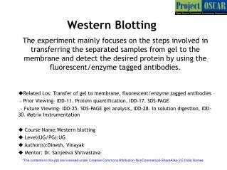
Introduction • Common techniques include: • Southern blotting (DNA) , • Northern blotting (RNA) , • and immunoblotting (for protein; also known as Western blotting).
1- Electrophoresis 2- Transfer 3- Blocking • The blotting procedures can be divided into six main steps 4- Probing 5- Detection 6- Results
1- Electrophoresis • The molecule of interest is present in a complex mixture of molecules. • Separate on the basis of size. • Separating the molecules by gel electrophoresis on either: • an agarose • or polyacrylamide gel.
2- Transfer (blotting) • Following separation, the molecules are transferred to a solid support such as: • a nylon, • nitrocellulose, • or polyvinylidene fluoride (PVDF) membrane. • Carbon copy of the molecules that were present in the gel are now immobilized on a membrane.
Type of Membrane The membrane of Choice The membrane of choice is determined by the sensitivity required and the detection method to be used.
Agar gel with DNA Weight Wick (filter paper) Filter paper Buffer Membrane 2- Transfer (blotting)- Capillary Transfer • Fragments are eluted from the gel and deposited onto the membrane by buffer that is drawn through the gel by capillary action. Paper towel stack
Whatman paper - Nitrocellulose filter + Buffer Buffer Gel Glass plates 2- Transfer (blotting)- Electrophoretic Transfer • The negatively charged nucleic acid molecules will move from the gel to the membrane
2- Transfer (blotting)- Vacuum Transfer • Nucleic acids are eluted by buffer that is drawn through the gel by application of negative pressure (a vacuum). Gel Recirculating buffer Nitrocellulose filter Vacuum Porous plate
2- Transfer (blotting)- Cross-Linking • Once transferred to a membrane, they have to be linked to the membrane. • UV irradiation, covalently attach the nucleic acids to the membrane • Covalent bond between the amide groups on the nylon and the carbonyl groups found on the thymine and uracil bases
2- Transfer (blotting)-Cross-Linking • Alternatively, the membrane can be baked at 80◦C for 2 hr. • Dehydration of the nucleic acids on the blot, • resulting in the generation of stable hydrophobic interactions between the nucleic acid and the membrane.
3- Prehybridization (Blocking) • The transferred nucleic acids only occupy a limited amount of the surface area of the membrane. • The molecules in the prehybridization solution coat the rest of the membrane. • In the absence of such a treatment, the probe would: • associate with the unoccupied sites on the membrane, • resulting in very high background and a very low signal-to-noise ratio.
4- Probing • Membrane is now incubated with a specific probe that binds to the protein or nucleic acid sequence of interest. • For southern or northern, a fragment of DNA of variable length (usually 100-1000 bases long)
4- Probing • The probe will have two properties: • First, anneal specifically with the sequence of interest. • Second, modified in such a way as to allow for the detection of the annealed sequences.
4- Probing • Probe used for an immunoblot is an antibody that recognizes a particular protein • 2o Ab with a label will bind to 1o Ab with high affinity • Unbound probe or nonspecifically bound probe is removed by washing the membrane
4- Probing Probe
4- Probing- Production of Probes • The availability of a gene probe is essential in many molecular biology techniques. • The information needed to produce a gene probe may come from many sources, • e.g. genetic databases. • Genbank and EMBL search to identify particular sequences relating to a specific gene or protein.
4- Probing- Production of Probes • Use related proteins from the same gene family to gain information DNA sequence. • Similar proteins or DNA sequences but from different species may also provide a starting point with which to produce a probe.
4- Probing- Labeling of Probes • To visualize DNA or RNA, the nucleic acid should to be attached to a label: • radioactive, • colored, • fluorescent, • Or luminescent. • The three main choices are: • radioisotopes, • fluorophores, • and small-molecule binding partners
4- Probing- Labeling of Probes- A-Radioisotopes • 32P is commonly used as a label • Emits radiation that can be easily detected by autoradiography • Nucleotides that incorporate 32P are commercially available. • Can be readily incorporated into DNA by enzyme-catalyzed reactions.
4- Probing- Labeling of Probes- B- Fluorophores • Fluorophores are molecules that absorb light at one wavelength and then emit light at a different wavelength. • Incorporate fluorophores: • chemically during DNA synthesis, • or enzymatically
4- Probing- Labeling of Probes-C- Small molecule binding partners • Small organic molecules that are recognized by: • antibodies or • other protein binding partners. • Common molecules are biotin and digoxigenin
5- Detection • Streptavidin covalently conjugated with a detection moiety. • For example, streptavidin conjugated directly to a fluorophore or to enzymes such as horseradish peroxidase or alkaline phosphatase. • Enzymes detected by their action on provided substrates that deposit products which are colored, luminescent, or fluorescent.
4- Probing- Labeling of Probes-C- Small molecule binding partners
Hybridization • Probe is generated and added to the blot for 1 to 24 hr. • Time to hybridize the blot depends on a variety of factors and must be determined empirically. • Overnight to maximize hybridization of the probe to the target
5- Detection • To visualize the bound probe. • Determined by the nature of the probe. • If a radioactive probe, autoradiography • exposure of the blot to X-ray film will allow for detection and quantitation of the bound probe.
5- Detection • If chemical- or enzyme-based, • substrates are added • the resulting signal is developed • and can be documented by: • colorimetric, • or chemiluminescent imaging.
5- Detection • For fluorescently labeled nucleic acid • use imaging equipment to excite the fluorophore • And the appropriate filter to detect the emitted light.
6- Results and Analysis • Once the blot is developed, the resulting banding pattern can be analyzed. • Analysis involves: • determining the amount and molecular weight or size of the molecules on the blot • and comparing the results to the predicted pattern. • To determine the molecular weight a standard curve of size versus migration distance is derived from the molecular weight markers
3 Best Fit Line 2 Log- Molecular Weight 1 Distance (mm)
Positive & Negative Controls Negative controls • Include samples that are identical to the experimental sample but are missing the target that the probe is supposed to recognize. • Very useful in determining the existence of any background that can be due to cross-reactivity between the probe and the sample.
Positive & Negative Controls Positive control • Include samples that contain the protein or nucleic acid of interest. • When included in the experiment allows the investigator to confirm that the experiment was successfully executed. • No signal indicates that the problem lies with the experimental samples and not with the procedure.
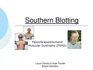
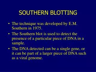
Southern Blotting • Developed by E.M. Southern in 1975. • A technique used in molecular biology to check for the presence of a particular DNA sequence in a DNA sample.
Flow chart of Southern hybridization Preparing the samples and running the gel Southern transfer & Fixing DNA onto membrane Probe preparation Prehybridization Hybridization Post-hybridization washing Signal detection Isotope Non-isotope
Preparing the samples and running the gel • Extraction of DNA • DNA must first be fragmented into small pieces that can migrate through an agarose gel matrix. • Restriction enzymes are used to fragment the DNA
Preparing the samples and running the gelDNA Digestion • Restriction enzymes recognize specific DNA sequences in DNA and cleave the DNA at these restriction sites. • Digestion with a given restriction enzyme produces a set of fragments that are easily separated by agarose gel electrophoresis.
Preparing the samples and running the gelDNA Digestion The enzyme EcoRI cutting DNA at its recognition sequence
Preparing the samples and running the gelElectrophoresis • Nucleic acids are negatively charged at a neutral pH • This allows their migration through an electric field • Agarose is a highly porous polysaccharide that acts as a sieve, allowing the fragments of DNA to be separated according to length.
Preparing the samples and running the gelDenature the DNA • Denature DNA with an alkaline solution such as NaOH. • Double stranded becomes single-stranded. • Single strands are ready to be transferred to a solid support
Southern Transfer & Fixing DNA • Transfer the DNA from the gel to a solid support. • Baking the membrane at 80°C for 2 h in a vacuum oven. • Or expose to ultraviolet
Probe Preparation, Prehybridization & Hybridization • A labeled probe is prepared which is complementary for the sequence we are looking for • Prehybridization to block sites where probe can bind on the membrane • Hybridization
Post-hybridization washing • Following hybridization, the blot must be washed to remove unassociated and nonspecifically annealed probe from the blot.