Download
1 / 78
780 likes | 1.02k Vues
Chase Findley, MSIV. USMLE STEP I Review Week 1: Cell Bio & Histology. Basic Cell Biology. Cell Cycle Phases. Checkpoints control transitions between cell phases. Regulated by cyclins, cdks, and tumor suppressors. Cell Cycle Phases. Permanent cells Remain in G0, regenerate from stem cells

E N D
Chase Findley, MSIV USMLE STEP I Review Week 1: Cell Bio & Histology
Cell Cycle Phases • Checkpoints control transitions between cell phases. Regulated by cyclins, cdks, and tumor suppressors.
Cell Cycle Phases • Permanent cells • Remain in G0, regenerate from stem cells • Neurons, skeletal and cardiac muscle, RBC’s • Stable cells • Enter G1 from G0 when stimulated • Hepatocytes, lymphocytes • Labile cells • Never go to G0, divide rapidly with short G1 • Bone marrow, gut epithelium, skin, hair follicles
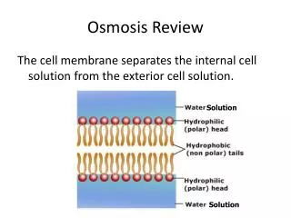
Plasma Membrane Composition • Asymmetric fluid bi-layer • 50% cholesterol, 50% phospholipids • Small amounts of protein, sphingolipids, glycolipids • High cholesterol or long saturated fatty acid content increases melting temperature
Endoplasmic Reticulum • Rough • Site of synthesis of secretory (exported) proteins and N-linked oligosaccharide addition • In neurons, (Nissl bodies) synthesize enzymes and peptide neurotransmitters • Mucous secreting goblet cells and antibody secreting plasma cells are rich in RER
Endoplasmic Reticulum • Smooth • Site of synthesis of steroids • Detoxification of drugs and poisons • Liver hepatocytes and adrenal cortex are rich in SER
Golgi Apparatus • “Distribution center” of proteins and lipids from ER to plasma membrane, lysosomes, secretory vesicles • Adds mannose-6-phosphate to proteins, targeting to lysosome • Failure results in I-cell disease, enzymes secreted outside cell • Proteoglycan assembly and sulfation
Microtubules • Helical array of polymerized dimers of α and β tubulin • Each dimer has 2 GTP bound • Incorporated into flagella, cilia, mitotic spindles, neurons • Chediak-Higashi syndrome • Defect in microtubule polymerization with decreased phagocytosis • Target of mebendazole, taxol, griseofulvin, vincristine, vinblastine, colchicine
Cilia Structure • 9+2 arrangement of microtubules • Dynein (ATPase) links peripheral 9 doublets, causes bending by differential sliding of doublets • Dynein=retrograde Kinesis=anterograde • Kartagener’s syndrome • Dynein defect, immotile cilia, infertility, recurrent infections
Collagen • Most abundant protein in body • Organizes, strengthens extracellular matrix • Type I • Bone, skin, tendon, dentin, fascia, cornea • Type II • Cartilage, vitreous body, nucleus pulposus • Type III (Reticulin) • Skin, blood vessels, uterus, fetal tissue • Type IV • Basement membrane
Collagen Synthesis • Inside fibroblasts • Synthesis (RER) • Translation of collagen α-chains (preprocollagen) • Hydroxylation (ER) • Specific proline and lysine residues, requires Vitamin C • Glycosylation (Golgi) • Pro-α chain residues, formation of procollagen (triple helix of α-chains) • Exocytosis • Procollagen exocytosed to extracellular space
Collagen Synthesis • Outside fibroblasts • Proteolytic processing • Cleavage of terminal regions of procollagen, transforms into insoluble tropocollagen • Cross-linking • Reinforcement of many staggered tropocollagen molecules by covalent lysine-hydroxylysine cross-linkage, produces collagen fibrils • Defective collagen synthesis causes Ehlers-Danlos syndrome.
Elastin • “Stretchy” protein • Rich in proline, lysine • Found in lungs, large arteries, elastic ligaments • α-1 antitrypsin inhibits elastase, excessive elastase activity causes emphysema
Phosphotidylcholine (Lecithin) Function • Major component of RBC membranes, surfactant, myelin, bile • Used in esterification of cholesterol
Immunohistochemical Stains • Connective Tissue • Muscle • Epithelial Cells • Neurons • Neuroglia • Vimentin • Desmin • Cytokeratin • Neurofilaments • Glial fibrillary acid proteins
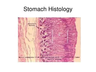
Digestive Tract Histology • Mucosa • Contains epithelium, lamina propria, muscularis mucosa • Absorptive function, villae • Submucosa • Contains submucosal nerve plexus • Muscularis externa • Contains Myenteric nerve plexus • Inner circular, outer longitudinal • Serosa/adventitia
Digestive Tract Histology • Submucosal nerve plexi • Submucosal layer • Coordinates secretions, blood flow, absorption • Myenteric nerve plexi • Muscularis externa layer • Coordinates motility
Digestive Tract Histology • Brunner’s Glands • Located in duodenal submucosa • Secrete alkaline mucous, neutralize acidic stomach contents • Hypertrophy in peptic ulcer disease
Digestive Tract Histology • Peyer’s Patches • Unencapsulated lymph tissue in mucosa and submucosa of small intestine • Take up antigen, stimulate local B cells to differentiate into IgA-secreting plasma cells • IgA secreted into lumen
Digestive Tract Histology • Barrett’s Esophagus • Replacement of non-keratinized, squamous epithelium with intestinal columnar epithelium in distal esophagus • Caused by acid reflux, may lead to adenocarcinomas • Example of metaplasia
Liver Histology • Zone 1 • Periportal • Sensitive to toxicinjury • Zone 2 • intermediate • Zone 3 • Pericentral • Sensitive to ischemic injury
GI Secretory Cells (More thoroughly covered in GI session) • Parietal Cells (Stomach) • Intrinsic factor • B12 absorption, destroyed in pernicious anemia • Gastric acid (HCl) • Chief Cells • Pepsin • Protein digestion • Mucosal Cells • Bicarbonate • G Cells • Gastrin
Erythrocytes • Anucleate • Biconcave • High surface area to volume ratio for easy gas exchange • Life span: 120 days • Glucose energy source • 90% anaerobically degraded to lactate • Membrane contains chloride-bicarbonate antiport, involved in “physiologic chloride shift”
Erythrocytes • Anisocytosis • Varying size • Poikilocytosis • Varying shape • Reticulocyte • Immature erythrocyte • Larger, bluish tinge
Neutrophils • Multilobed nucleus • Mediate acute inflammatory response • Phagocytic • Primary granules contain hydrolytic enzymes, lysozyme, myeloperoxidase • Hypersegmented in B12/folate deficiency
Neutrophils • Normal • Hypersegmented
Leukocytes • Granulocytes • Basophils, eosinophils, neutrophils • Mononuclear cells • Lymphocytes, monocytes
Lymphocytes • Round, densely staining nucleus • Little cytoplasm • T & B lymphocytes
T Lymphocytes • Mediate cellular immune response • Originate from stem cells in bone marrow, mature in thymus • Differentiate into: • Cytotoxic T cells • MHC I, CD8 • Helper T cells • MHC II, CD4 • Suppressor T Cells
B Lymphocytes • Mediate humoral immune response • Originate from stem cells in bone marrow, mature in marrow • Migrate to peripheral lymph tissue • Differentiate into plasma cells, produce antibody when presented with antigen • Function as APC via MHC II
Mast Cells • Mediate allergic reaction • Contain histamine, heparin, chemotactic factors • Bind IgE to cell membrane • Found in tissue • Cromolyn sodium prevents degranulation
Monocytes • Kidney shaped nucleus • Differentiates to macrophages in tissue
Macrophages • Phagocytic for bacteria, cell debris, senescent blood cells • Activated by gamma interferon • Function as antigen presenting cell via MHC II
Plasma Cells • Off-center nucleus, clock-face chromatin • Abundant rough endoplasmic reticulum and Golgi apparatus • Differentiate from B cells, produce antibody
Eosinophils • Bilobate nucleus • Highly phagocytic for antigen-antibody complexes • Defend against helminth and protozoan infections • Elevated in allergies, asthma certain neoplasms, collagen vascular diseases
Basophils • Bilobate nucleus • Mediate allergic reaction • Contain histamine, heparin, leukotrienes • Found in blood
Epidermal Layers *Langerhan’s cells are dendritic cells that function as APC’s in skin. Remember Birbeck granules!
Epithelial Cell Junctions • Zona occludens (tight junction) • Creates semi-permeable barrier • Macula adherens • Small discrete points of attachment • Gap junction • Allows adjacent cells to communicate via metabolic/electrical processes • Hemidesmosome • Anchors cells to extracellular matrix • Integrin • Maintains integrity of basement membrane
Skeletal Muscle Cell Structure • Sarcomere • Skeletal muscle unit from Z line to Z line • A band • Area of overlap of actin and myosin • I band • Area of actin only Contraction causes I band shortening, A band stays same