Download
1 / 22
220 likes | 225 Vues
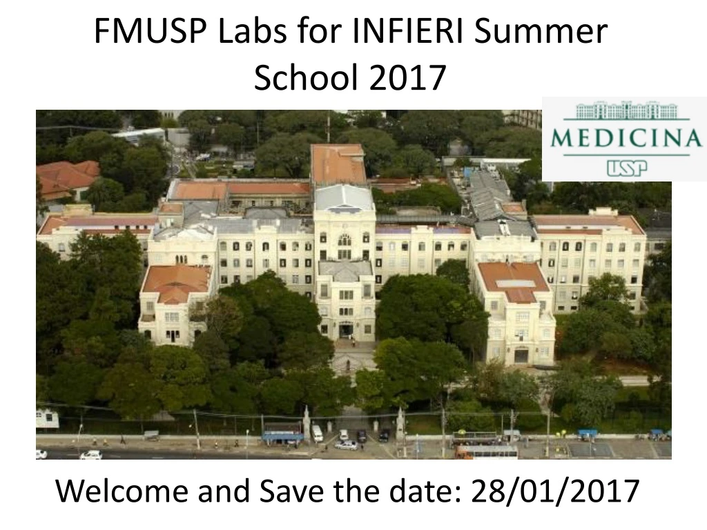
FMUSP Labs for INFIERI Summer School 2017. Welcome and Save the date: 28/01/2017. Hospital das Clínicas HC-FMUSP. Main objectives of our labs. Allow you to get to know different research facilities in Medical Imaging at FMUSP Basic concepts of the different equipements (hardware)

E N D
FMUSP Labs for INFIERI Summer School 2017 Welcome and Save the date: 28/01/2017
Main objectives of our labs • Allow you to get to know different research facilities in Medical Imaging at FMUSP • Basic concepts of the different equipements (hardware) • Basic concepts of image formation (contrast, signal to noise ratio, resolution)
Medical Imaging: MRI lab session Where: PISA core facility at FMUSP with 7T whole body scanner KhallilChaim Maria Otaduy
Larmor frequency (resonance) w0= g. B0 ÁguaLivre MRI do Joelho @7T MRI BASIC PRINCIPLES AND APPLICATIONS, 3ed., Mark A. Brown & Richard C. Semelka Journal of Magnetic Resonance Imaging Volume 30, Issue 3, pages 606-614, 26 AUG 2009 DOI: 10.1002/jmri.21881
High magnetic field is not all advantages, we have big challenges: strong SAR effects and strong dielectric effects. This implies in different hardware strategies for high field MRI (i.e. parallel transmission)
MRI lab tasks • 1st task: after listening to some instructions you will have to answer some questions about MRI safety • 2nd: After positioning MRI resolution phantom you will have to change image resolution (FOV, matrix and slice thickness) • 3rd task: After positioning contrast phantom (different T1 and T2) you will have to observe how the image changes with TE, TR and with echo acquisition mode (gradient-echo vs. spin-echo) • 4th task: After positioning MRS phantom you will have to describe how signal changes from time domain to frequency domain. Observe important characteristics of the final signal, and how does this signal change with a good B0 shimming (you will have to try out different shimming techniques).
Medical Imaging: PET lab session Where: Nuclear Medicine unit at InRad PET/MRI (3T) scanner AlexandreGarcez Bruno Pastorello
PET basic steps Ziegler S, Nuclear Physics, 2005 Biological radioactive nuclei: oxygen-15, fluorine-18, carbon-11, or nitrogen-13 Most used: 18-fluorodeoxyglucose (FDG) for glucose consumption, which is higher in tumors A PET scan measures also important body functions, such as blood flow, oxygen use, and sugar (glucose) metabolism.
PET/MRI labtasks • 1st task: After accompanying the calibration process of the equipment, list 4 calibration parameters and indicate the purpose of the calibration. • 2nd task: After observing how the phantom is filled, state the purpose of having spheres of different diameters. • 3rd task: After reconstructing the images using several parameters, describe what you observed in relation to the quality of the images. • 4th task: After analyzing the phantom images, list your results in terms of the contrast of the "hot" and "cold" spheres and compare with the values published by the manufacturer.
Do not worry with radioactivity!! • There will be no exposure for students • For some procedures a simulator will be used to mock the real process
Medical Imaging: Ultrasound Lab Where: InRad, Portaria 6 (convênio). MateusAranha Eugenio Martinetto
US: interaction with sound wavespenetration, attenuation and reflection • As sound penetrates all tissues, the signal becomes attenuated. • The further the sound penetrates, the greater the attenuation (objects far away are harder to see) • Reflection is required for the US probe to receive a signal to analyze (no reflection, no signal)
1st task: How looks US signal on phantom belowDifferent tissues have different degrees of penetration, attenuation and reflection (US contrast)
2nd task: What happens changing frequency, probe and angulation?
3rd task: Identify possible sources of artifact • (refraction, shadowing, enhancement, reverberation, and mirroring) • 4th task: Describe Doppler-effect and its clinical use in US
MRI Meeting point: Portaria 1 InRad US PET
Meeting point: Portaria 1 InRad
Meeting logistics • Hospital environment. Security controls. Needs to register one day in advance. • Until 14.00 at meeting point: Portaria 1 InRad (Institute of Radiology)-Av. Dr. Enéas de CarvalhoAguiar 255. • Direct contact with Lab Organizers: • MRI 7T maria.otaduy@hc.fm.usp.br or khallil.chaim@hc.fm.usp.br • US mateus.aranha95@gmail.com or emarinetto@hggm.es • PET alexandre.garcez@hc.fm.usp.br or bruno.pastorello@hc.fm.usp.br