Download

1 / 26
270 likes | 621 Vues
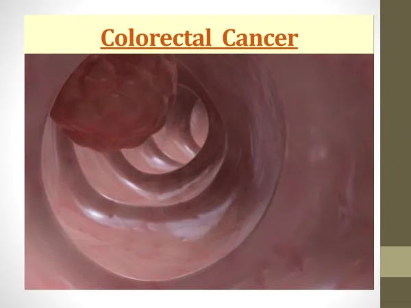
Colorectal Cancer. Colorectal Cancer. Worldwide, colon and rectum cancer is the third most common cancer and it is the most common GI cancer. 1. 2.

E N D
Colorectal Cancer Worldwide, colon and rectum cancer is the third most common cancer and it is the most common GI cancer. 1 2 The vast majority of colorectal cancers are adenocarcinomas, which arise from preexisting adenomatous polyps that develop in the normal colonic mucosa. Of patients with colon cancer, 90% are older than 50 years. The highest rates of incidence are in individuals aged 70-85 years. Only 5% of patients are younger than 40 years. 3 4 The overall 5-year survival rate from colon cancer is approximately 60% but it is different for each stage. For Dukes stage A tumors involving only the mucosa, the 5-year survival rate exceeds 90%, whereas for metastatic colon cancer, the 5-year survival rate is about 5%.
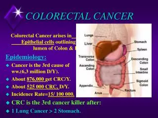
Colorectal Cancer Colon and rectum cancers accounted for about 1 million new cases in 2002 In the US: Colorectal cancer is the third most common malignant neoplasm worldwideand the second leading cause of cancer deaths in the United States. It is estimated that there will be 145,290 new cases diagnosed in the United States in 2005 and 56,290 deaths due to this disease. The highest incidence rates are in North America, Australia/New Zealand, Western Europe, and, in men especially, Japan. Incidence tends to be low in Africa and Asia and intermediate in Eastern Europe and southern parts of South America. From: Global Cancer Statistics, 2002 -- Parkin et al_ 55 (2) 74 -- CA A Cancer Journal for Clinicians
Colorectal Cancer Sex: The frequency of colon cancer is essentially the same among men and women.
Colorectal Cancer Spatial Distribution: • The most common sites are the rectum (34%) and sigmoid (25%). • Over the 20 years, the incidence of cancer in the cecum increased and that in the rectum decreased. • Cecal, ascending, and transverse colon cancers accounted for 34% of lesions - all beyond the range of the flexible sigmoidoscope. ClinicalPresentation: • Colon cancer often is found by screening and may be completely asymptomatic. • Approximately 50% of patients present with abdominal pain, • 35% with altered bowel habits, • 30% with occult bleeding, • 15% with intestinal obstruction. • Right-sided colon cancers tend to be larger and more likely to bleed, whereas left-sided tumors tend to be smaller and more likely to be obstructing.
Colorectal Cancer • Preferred Examination: • Begin the evaluation with a history and physical examination,including a digital rectal examination. • Inspect the stool and test for occult blood. • Perform blood tests, including a full blood count, liver function tests, and carcinoembryonic antigen level. • Perform either a sigmoidoscopy (rigid or flexible) and a double-contrast barium enema or a colonoscopy • Virtual colonoscopy, a new experimental test to evaluate the entire colon Double-contrast barium enemas are an option for screening for colorectal cancer and can aid in establishing the diagnosis of colon cancer. Double-contrast barium enema detects approximately 90% of colonic tumors.
Colorectal Cancer • Double-contrast study: • Most colonic cancers are relatively advanced, measuring 3-4 cm in diameter at diagnosis. The appearances of the tumors on double-contrast barium enema reflect the 3 morphologic types: polypoid, annular, or flat. • Polypoid lesions vary from small smooth tumors to larger lobulated masses with an irregular surface and an associated contour deformity along 1 margin of the bowel wall. • Annular lesions result from irregular circumferential masses that severely constrict the bowel lumen. The margins of the carcinoma show overhanging edges, the tumor shelf or shoulder (termed "apple-core" lesion). The mucosal folds in the narrowed segment are destroyed; ulceration may be present • Flat lesions, which are rare, are visualized as a unilateral broad-based contour defect. Ulceration may be present
Colorectal Cancer Double-contrast study: Polypoid carcinoma. A large, irregular lobulated mass is present in the rectosigmoid junction. From: http://www.kgan.minami.fukuoka.jp
Colorectal Cancer Double-contrast study: Typical annular carcinomaof the transverse colon From: http://www.kgan.minami.fukuoka.jp
Colorectal Cancer Double-contrast study: "apple-core" lesion Annular carcinoma of the sigmoid colon. The lumen of the sigmoid is narrowed severely by the circumferential mass with mucosal destruction and the overhanging edges or shouldering at the tumor margins.
Colorectal Cancer Double-contrast study: Flat carcinoma of the sigmoid colon - a unilateral broad-based contour defect. From: http://www.kgan.minami.fukuoka.jp
Colorectal Cancer CAT SCAN • Indications for CT scan • CT scan is used for staging colonic carcinoma prior to surgery, for assessing and staging recurrent disease, and for detecting the presence of distant metastases. • Preoperative CT scan is indicated if distant metastases or local invasion of adjacent organs or abdominal wall are suggested clinically. • In older patients who may be unable to undergo colonoscopy or barium enema, modified CT scan may be performed for primary detection of colorectal tumors. • Colonic tumors may be diagnosed on CT scan as an incidental finding.
Colorectal Cancer • CT Findings • A localized tumor may be seen on CT scan as an intraluminal or intramural mass of soft tissue density adjacent to the gas-filled or contrast-filled bowel lumen. • More advanced tumors are associated with thickening of the bowel wall (>6 mm) and infiltration of the pericolic fat. • Annular carcinomas are detected by a thickening of the bowel wall and narrowing of the lumen. This thickening is concentric if the scanning plane is at right angles to the long axis of the bowel. • Extracolonic tumor spread is indicated by a loss of tissue fat planes between the colon and surrounding structures • Tumors less than 2 cm in diameter cannot be detected reliably by the standard CT scan technique.
Colorectal Cancer CT Scan Staging System For Colonic Cancer modified from Thoeni N staging Nodes greater than 10 mm in diameter are considered abnormal. M Staging Hepatic metastases are the most common site of distant spread. Other common sites include the lungs, adrenals, peritoneum, and omentum.
Colorectal Cancer CT Findings Preoperative CT – colon wall thickening and infiltration of the pericolic fat
Colorectal Cancer CT Findings Numerous metastases. The tumor cells were arranged in nodules and occupied approximately 90% of the hepatic parenchyma. Contrast-enhanced CT showing liver metastases. Several low-density metastasesinvolve both lobes of the liver.
Colorectal Cancer CT Findings – CT colonography • CT scan colonography or virtual colonoscopy was introduced in 1996 as a screening tool for the detection of colorectal polyps and small cancers. • It involves a 3-dimensional computer reconstruction from a volumetric data set using a workstation as well as distending a clean colon with air. • Images are read as soft copy from the workstation using a combination of paging-through the 2D axial images, aided by multiplanar and 3D endoluminal images. The sensitivity of this technique is greater than that of the double-contrast barium enema. For polyps larger than 10 mm, it has a sensitivity of 91% but a specificity of 76%. This sensitivity falls to 81% for 5-10 mm polyps.
Colorectal Cancer CT Findings – CT colonography The recent arrival of multisectional helical scanners has reduced the time required to obtain the images (usually 30 seconds for each series, scanning the patient prone and supine using a reduced tube current to minimize the radiation dose).
Colorectal Cancer CT Findings – CT colonography
Colorectal Cancer CT Findings – CT colonography
Colorectal Cancer Ultrasound • The primary role of ultrasound (US) in patients with colonic cancer is the detection of hepatic metastases. • US has a detection rate of 70-90% for hepatic metastases.
Colorectal Cancer MRI • MRI provides greater contrast between soft tissues than CT scan. • Colonic tumors have low signal intensity (similar to adjacent skeletal muscle) on T1-weighted sequences, which facilitates their differentiation from high-signal perirectal fat. • T2-weighted images are used to detect pelvic sidewall invasion. • MRI and CT scan have a similar overall accuracy (approximately 60%) in the detection of enlarged lymph nodes (N staging) and liver metastases (M staging). • MRI has a higher sensitivity (91%) than CT scan (82%) in detecting local recurrence and a higher specificity (100%) than CT scan (69%).
Colorectal Cancer PET • Findings: • Nuclear medicine has a small peripheral role in colonic cancer. • Consider using radioimmunoscintigraphy with monoclonal antibody that recognizes carcinoembryonic antigen (CAA) or tumor-associated glycoprotein-72 to detect disease recurrence in the pelvis or extrahepatic abdomen. • Consider using positive emission tomography (PET) with 2F 18-fluoro-deoxy-D-glucose (FDG) to detect recurrent disease.
Colorectal Cancer PET/CT HistoryA 70-year-old male with hx of colorectal CA s/p resection, chemo and XRT. Recently with rising CEA levels and CT with indistinguishable soft tissue density in pelvis. PET/CT FindingsUptake in soft-tissue mass plus an additional metastatic lesion in sacrum. From: http://www.petscaninfo.com/
Colorectal Cancer - Screening Prevention and Screening Methods 1.5 cm bi-lobed benign tubular adenoma on a stalk. • Between 70 and 90 percent of colorectal cancers arise from adenomatous polyps, and 10 to 30 percent arise from sessile adenomas. • The larger the polyp, the greater the potential for malignancy. • Diminutive polyps (5 mm or less in diameter) have a negligible malignant potential. • Polyps with a diameter of 5 to 10 mm are considered small, whereas polyps greater than 10 mm in diameter are considered large. • Polyps larger than 2 cm in diameter have a 50 percent chance of becoming malignant over time. Sessile villous adenoma