Download
1 / 72
760 likes | 1.33k Vues
Down Syndrome (Trisomy 21 ( Trisomy 13 & 18. Dr Pupak Derakhshandeh, PhD Ass Prof Medical Science of Tehran University. What are chromosomes?. Chromosomes are the structures that hold our genes Genes are the individual instructions that tell our bodies how to develop and function

E N D
Down Syndrome(Trisomy 21(Trisomy 13 & 18 Dr Pupak Derakhshandeh, PhD Ass Prof Medical Science of Tehran University
What are chromosomes? Chromosomes are the structures that hold our genes Genes are the individual instructions that tell our bodies how to develop and function They govern our physical and medical characteristics, such as hair color, blood type and susceptability to disease. Each chromosome has a p and q arm; p is the shorter arm and q is the longer arm. The arms are separated by a pinched region known as the centromere
How many chromosomes do humans have? • The typical number of chromosomes in a human cell is 46 - two pairs of 22 + XX/XY • Holding an estimated 30,000 to 35,000 genes. • One set of 23 chromosomes is inherited from the biological mother (from the egg), and the other set is inherited from the biological father (from the sperm).
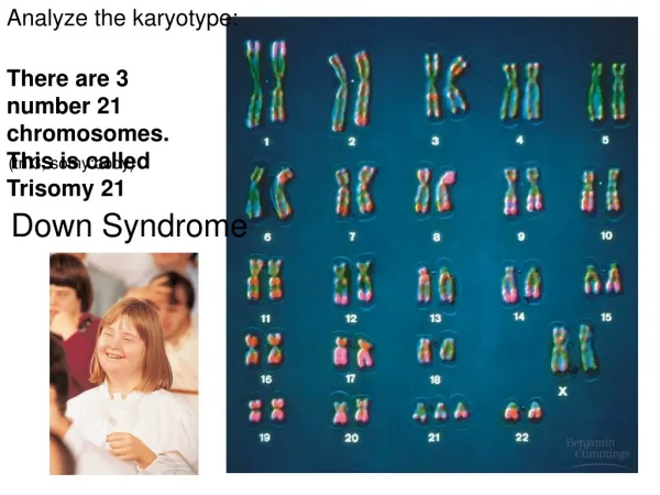
study of the chromosomes • with a microscope, then Stainning • The chromosomes look like strings with light and dark "bands" • A picture, or chromosome map, of all 46 chromosomes is called a karyotype • The karyotype can help identify chromosome abnormalities that are evident in either the structure or the number of chromosomes.
The pairs have been numbered from 1 to 22, with the 23rd pair labeled "X" and "Y." • In addition, each chromosome arm is defined further by numbering the bands that appear after staining • The higher the number, the further that area is from the centromere. • The first 22 pairs of chromosomes are called "autosomes" • Final pair is called the "sex chromosomes." • The sex chromosomes an individual has determines that person's gender; females have two X chromosomes (XX), and males have an X and a Y chromosome (XY).
How Chromosome Abnormalities Happen? Meiosis Mitosis Maternal Age Environment
Meiosis • Chromosome abnormalities : • happen as a result of an error in cell division. “Meiosis” is the name used to describe the cell division that the egg and sperm go through when they are developing. • Normally, meiosis causes a halving of chromosome material, so that each parent gives 23 chromosomes to a pregnancy
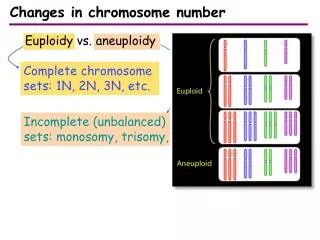
Chromosome abnormalities • Abnormality of chromosome number or structure: • Numerical Abnormalities • Structural Abnormalities
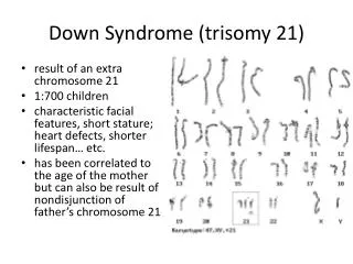
Numerical Abnormalities • When an individual is missing either a chromosome from a pair (monosomy) or has more than two chromosomes of a pair (trisomy). • An example: Down Syndrome, also known as Trisomy 21 (an individual with Down Syndrome has three copies of chromosome 21, rather than two).
Numerical Abnormalities • Kleinfelter Syndrome is an example of trisomy the individual is born with three sex chromosome, XXY. • Turner Syndrome is an example of monosomy the individual is born with only one sex chromosome, an X.
Down Syndrome (Trisomy 21( Trisomy 2(
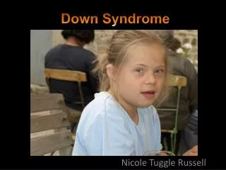
Down syndrom) Trisomy 21, 47) critical region: • A region on the long (q) arm of chromosome 21 • Down syndrome causes mental retardation • a characteristic facial appearance • multiple malformations
critical region: • Associated with a major risk for heart malformations • a small but still significant risk of acute leukemia • 3 copies of chromosome number 21
incidence of 1 in 660 and is by far the most common chromosomal abnormality Slight flattening of the face • A low bridge of the nose (lower than the usually flat nasal bridge of the normal newborn) • An epicanthal fold (a fold of skin over top of the inner corner of the eye, which can also be seen less frequently in normal babies) • A ring of tiny harmlesswhite spots around the iris • mental retardation
Down Syndrome: Prenatal Risk • The risk of trisomy 21 is directly related to maternal age • Patients who will be 35 years or older on their due date should be offered chorionic villus sampling or second-trimester amniocentesis
Women younger than 35 years should be offered maternal serum screening at 16 to 18 weeks of gestation • The maternal serum markers used to screen for trisomy 21 are alpha-fetoprotein, unconjugated estriol and human chorionic gonadotropin
The use of ultrasound to estimate gestational age improves the sensitivity and specificity of maternal serum screening. (Am Fam Physician 2000;62:825-32,837-8.)
Etiology and Clinical Manifestations • Trisomy 21 is present in 95 percent of persons with Down syndrome. • Mosaicism, a mixture of normal diploid and trisomy 21 cells, occurs in 2 percent.
Etiology and Clinical Manifestations • The remaining 3 percent have a Robertsonian translocation in which all or part of an extra chromosome 21 is fused with another chromosome.
Robertsonian translocation • The reciprocal transfer of the long arms of two of the acrocentric chromosomes: 13, 14, 15, 21 or 22 • On rare occasions, other non-acrocentric chromosomes undergo Robertsonian translocation
Robertsonian translocation • a reciprocal transfer of the whole long or short arms close to the centromere • A relatively common Robertsonian translocation is between chromosome 14 and chromosome 21 • In meiosis, a trivalent is formed.
Balanced reciprocal translocation Balanced reciprocal translocation Balanced reciprocal translocation
Frequency of Dysmorphic Signs in Neonates with Trisomy 21 Dysmorphic signFrequency (%) Flat facial profile90 Poor Moro reflex85 Hypotonia80 Hyperflexibility of large joints80 Loose skin on back of neck80 Slanted palpebral fissures80
Frequency of Dysmorphic Signs in Neonates with Trisomy 21 Dysmorphic sign Frequency (%) Dysmorphic pelvis on radiograph70 Small round ears60 Hypoplasia of small finger, middle phalanx60 Single palmar crease45
Persons with Down syndrome usually have mild to moderate mental retardation • School-aged children with Down syndrome often have difficulty with language, communication • Adults with Down syndrome have a high prevalence of early Alzheimer's disease
Incidence of Some Associated Medical Complications in Persons with Down Syndrome DisorderIncidence (%) Mental retardation>95 Growth retardation>95 Early Alzheimer's disease 75% by age 60 Congenital heart defects (atrioventricular canal defect, ventricular septal defect, atrial septal defect40
DisorderIncidence (%) Hearing loss 40 to 75 Ophthalmic disorders (congenital cataracts, glaucoma( 60 Epilepsy5 to 10 Gastrointestinal malformations (duodenal atresia, Hirschsprung disease)5 Hypothyroidism5 Leukemia5
DisorderIncidence (%) Increased susceptibility to infection (pneumonia, otitis media, sinusitis, pharyngitis( 1-6 Infertility>99% in men anovulation in 30% of women
. . . Estimated risk of Down syndrome according to maternal age Maternal Serum Screening Maternal Serum Screening Maternal Serum Screening
The risk of having a child with Down syndrome • 1/1,300 for a 25-year-old woman; • at age 35, the risk increases to 1/365 • At age 45, the risk of a having a child with Down syndrome increases to 1/30
Maternal Serum Screening • If all pregnant women 35 years or older chose to have amniocentesis • about 30 percent of trisomy 21 pregnancies would be detected • Women younger than 35 years give birth to about 70 percent of infants with Down syndrome
The risk of having a child with Down syndrome • Maternal serum screening (multiple-marker screening) can allow the detection of trisomy 21 pregnancies in women in this younger age group.
Maternal Serum Screening"triple test" or "triple screen""Multiples of the Median (MoM)" • Alpha-fetoprotein (AFP) • unconjugated estriol • human chorionic gonadotropin (hCG) • the serum markers most widely used to screen for Down syndrome
"Multiples of the Median (MoM)" • AFP is produced in the yolk sac and fetal liver. • Unconjugated estriol and hCG are produced by the placenta. • The maternal serum levels of each of these proteins and of steroid hormones vary with the gestational age of the pregnancy.
"Multiples of the Median (MoM)" • With trisomy 21, second-trimester maternal serum levels of AFP and unconjugated estriol are about 25 percent lower than normal levels • maternal serum hCG is approximately two times higher than the normal hCG level