Download
1 / 21
210 likes | 341 Vues
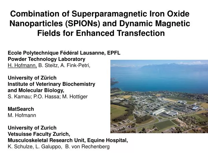
Combination of Superparamagnetic Iron Oxide Nanoparticles (SPIONs) and Dynamic Magnetic Fields for Enhanced Transfection. Ecole Polytechnique Fédéral Lausanne, EPFL Powder Technology Laboratory H. Hofmann, B. Steitz, A. Fink-Petri, University of Zürich

E N D
Combination of Superparamagnetic Iron Oxide Nanoparticles (SPIONs) and Dynamic Magnetic Fields for Enhanced Transfection Ecole Polytechnique Fédéral Lausanne, EPFL Powder Technology Laboratory H. Hofmann, B. Steitz, A. Fink-Petri, University of Zürich Institute of Veterinary Biochemistry and Molecular Biology, S. Kamau; P.O. Hassa; M. Hottiger MatSearch M. Hofmann University of Zurich Vetsuisse Faculty Zurich, Musculoskeletal Research Unit, Equine Hospital, K. Schulze, L. Galuppo, B. von Rechenberg
Aim of study • Explore the use of dynamic magnetic fields to enhance non-viral transfection with superparamagnetic iron oxide (SPION) beads • Uptake of SPION as non-viral vectors into cells (293T, COS and HeLa cell lines& synoviocytes) • Obtain preliminary results with intraarticular application of non-viral vector in vivo • Take advantage of • the smaller sized SPION-beads (70 nm) for intraarticular application • Lower cytotoxicity of PVA coated compared to PEI coated SPION
Important steps in transfection 1, 2 extracellular 3, 4 H+ 4 Early Endosome pH 6.5 – 7.5 Late Endosome pH ~ 5.0 Endocytosis intracellular GFP Endosomal Escape Nucleus Gene Products Transcription Nuclear Targeting
1 2 dB/dz Static field gradient Y Y Y Y Cell membrane Force per particle acting on the membrane : Static: 1 10-13 – 1 10-14 N Dynamic: 1 10-15 – 1 10-16 N 3 Y Y 4 Dynamic magnetic Field gradient
Materials & methods Transfection with SPIONs SPIONS:PVA-SPIONs, PEI-SPIONs (50 – 70 nm) ; Control: Larger SPIONs (200-250 nm) Cells: HeLa (human cervix carcinoma cells),293T, Cos7 (fibroblast , America Type Culture Collection (ATCC)), synoviocytes, Plasmid and PCR-Products: • pEGFP-C2 plasmid (Clontech) • PCR products from pEGFP-C2 plasmid (Clontech) were amplified using primers designed to include the 5’human cytomegalovirus (CMV) immediate early promoter and a 3’SV40 early mRNA polyadenylation signal. The PCR products (1.6 kilobase pairs) were purified using PCR purification Kit (Qiagen). Experimental procedure: SPIONs complexed with DNA for 30 min at room temperature Added to cells, exposure to static magnet for 5 or 20 min, 4 hrs incubation Medium replacement incubation for minimum 24 hrs FACS and microscopy analysis - 24 hrs (48, 72 and 96 hrs) Transfection controls: Lipofectamine, PEI and Ca2PO4 Detail see : Nucleic Acids Research2006 , accepted,
Magnetic flux density distribution (component Mz and Mx) z 30 y Mz M’z x Mag flux density (mT) M’x Mx 0 0 3 6 Distance (cm) Well plate Dynamic magnetic field generator Dynamic magnetic field (Equipment :Matsearch, Pully CH and Stetter Elektronik, D) mZ X
20nm PEI coated SPIONs R = PEI:Fe mass ratios
M12: amino-PVA Changes of the Zeta-potential with DNA adsorption
Uptake of amino-PVA-Cy3.5 in synoviocytes 300 24 h 48 h 72 h 24 hrs 82.1% 48 hrs 82.9% 72 hrs 85.8% 225 % of positive cells Cell count 150 75 0 10 0 10 1 10 2 10 3 10 4 Fluorescence intensity (Cy 3.5) Time (hours) Efficient uptake of PVA-coated SPIONs by synoviocytes
293T cells 293T cells Synovial cells 3.8% 96.2% 3.8% 96.2% Counts GFP GFP GFP Expression of GFP in cells transfected with PVA- and PEI-coated SPIONs PEI-SPIONs (24 hrs) PVA-SPIONs (96 hrs) • Efficient gene delivery by the SPIONs in different cells lines • PEI-SPIONs – high gene delivery after 24 hrs • PVA-SPIONs – clear gene expression after 72 hrs
293T cells were transfected with either DNA/polyMAG (A) or DNA/SPIONs (B),
Different cells were transfected with DNA/SPIONs (B), static magnetic field 5 or 20 min.
Enhancement of transfection rates by the application of dynamic magnetic fields 293T cells SPIONS + magnet • Application of dynamic magnetic field for 5 min significantly increased the transfection efficiency over that with the conventional transfection methods
Static and dynamic magnetic field bioweb.wku.edu/.../stokes/131f98chap8.html.
Behaviour of SPIONs in vivo after intraarticular application PEI-SPIONs 200-250 nm PEI-SPIONs 50 nm PVA-SPIONs 50 nm PEI-SPIONs 200-250 nm • Very good biocompatibility with PVA-SPIONs • Swelling, reddening, oedema with PEI-SPIONs especially with the larger particles • Immunohistochemistry for GFP identified positive samples in the inguinal lymphnode but was inconclusive for synovial membrane samples.
Summary • The uptake PVA-coated SPIONs is very efficient and they have better biocompatibility than PEI-SPIONs. • Significantly higher transfection rates were achieved with PEI-SPIONs after 5 or 20 min exposure to magnet and additionally 5 min to dynamic magnetic fields (than conventional transfection methods) • PEI-coated SPIONs are very efficient for non-viral gene delivery, resulting in high transfection rates especially with synoviocytes (96.2%) • Smaller PEI-SPIONs are more biocompatible than larger particles • In-vivo transfection with small PEI-SPION and dynamic magnetic fields seems to be possible.
Acknowledgement • Swiss National Science Foundation • VETSuisse Research Fund
Materials & methods Transfection with SPIONs SPIONS:PVA-SPIONs, PEI-SPIONs (50 – 70 nm) ; Control: Larger SPIONs (200-250 nm) Cells: 293T, HeLa, Cos7, synoviocytes, Plasmid and PCR-Products: • The pEGFP-C2 plasmid (Clontech) was propagated in Escherichia coli and purified using an Endotoxin-free Maxiprep plasmid Kit (Qiagen). • PCR products from pEGFP-C2 plasmid (Clontech) were amplified using primers designed to include the 5’human cytomegalovirus (CMV) immediate early promoter and a 3’SV40 early mRNA polyadenylation signal, (forward primer: 5’-CCG TAT TAC CGC CAT GCA T-3’; reverse primer: 5’-GCC GAT TTC GGC CTA TTG GT-3’). The PCR products (1.6 kilobase pairs) were purified using PCR purification Kit (Qiagen). Experimental procedure: SPIONs complexed with DNA for 30 min at room temperature Added to cells, exposure to static magnet for 5 or 20 min, 4 hrs incubation Medium replacement incubation for minimum 24 hrs FACS and microscopy analysis - 24 hrs (48, 72 and 96 hrs) Optimization: Aim: Efficient gene delivery (high proportion of green GFP expressing cells) Minimum toxicity (low proportion of red PI stained non-viable cells Transfection controls: Lipofectamine, PEI and Ca2PO4
PCR product promotor gene poly A side
Transfection with Plasmidic DNA vs. PCR product Plasmid Advantage • Large amounts are cheap • Stable • Large choice of transfection methods, promoters • Protocols optimized Disadvantage: • In vivo transfection regarded as non-safe PCR product Advantage: • In-vivo safe; without viral parts • No insertion into plasmid needed Disadvantage: • Expensive • Fast degredation Expression of unmodified PCR products never shown