Download
1 / 2
30 likes | 222 Vues
Diffusion MRI of Rodent Glioma at 21T Gregory S. Boebinger, National High Magnetic Field Laboratory DMR 0654118 NMR Spectroscopy and Imaging Facility User Program, Florida State University.

E N D
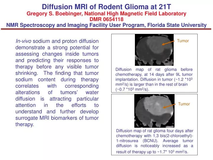
Diffusion MRI of Rodent Gliomaat 21TGregory S. Boebinger, National High Magnetic Field Laboratory DMR 0654118NMR Spectroscopy and Imaging Facility User Program, Florida State University In-vivo sodium and proton diffusion demonstrate a strong potential for assessing changes inside tumors and predicting their responses to therapy before any visible tumor shrinking. The finding that tumor sodium content during therapy correlates with corresponding alterations of tumors’ water diffusion is attracting particular attention in the efforts to understand and further develop surrogate MRI biomarkers of tumor therapy. Tumor Diffusion map of rat glioma before chemotherapy, at 14 days after 9L tumor implantation. Diffusion in tumor (~1.2 *103 mm2/s)is larger than in the rest of brain (~0.7 *103 mm2/s). Tumor Diffusion map of rat glioma four days after chemotherapy with 1,3 bis(2-chloroethyl)-1-nitrosurea (BCNU). Average tumor diffusion is noticeably increased as a result of therapy up to ~1.7* 103 mm2/s.
Sodium MRI of Rodent Gliomaat 21TGregory S. Boebinger, National High Magnetic Field Laboratory DMR 0654118NMR Spectroscopy and Imaging Facility User Program, Florida State University The first in vivo large rodent MR images were acquired using the NHMFL’s 21T MRI scanner. The high field experiments allowed a record high resolution for sodium and demonstrate a unique sensitivity of sodium MRI to tumor therapy. Sodium MRI and proton diffusion exhibit a strong correlation during tumor treatment. Both the high field and the capability of the simultaneous use of two imaging modalities for rodent glioma are valuable tools in evaluating tumor cellular changes and developing biomarkers of tumor’s responses to therapy. Reliable biomarkers are much needed for future development of efficient anti-tumor interventions. Tumor Sodium MRI of rat brain before chemotherapy. Tumor sodium content was elevated (~80 mM) relative to the rest of the brain (~50 mM). Resolution of sodium 3D image was 1µL, scan time was 30 min. Tumor Sodium MRI of rat brain four days after BCNU chemotherapy. Tumor sodium content increased as a result of therapy (~120 mM). Resolution of sodium 3D image was 1µL, scan time was 30 min.