Download
1 / 15
160 likes | 376 Vues
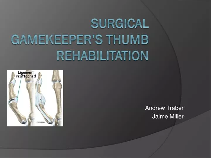
Andrew Traber Jaime Miller. Surgical gamekeeper’s thumb rehabilitation. Radiographs. What is gamekeepers thumb?. It’s a sprain to the thumb’s ulnar collateral ligament This use to occur when gamekeepers broke the neck’s of rabbits by using the thumb and index finger against the ground.

E N D
Andrew Traber Jaime Miller Surgical gamekeeper’s thumb rehabilitation
What is gamekeepers thumb? • It’s a sprain to the thumb’s ulnar collateral ligament • This use to occur when gamekeepers broke the neck’s of rabbits by using the thumb and index finger against the ground
Mechanism of Injury • Falling on an outstretched hand • Valgus force on the thumb while the thumb is abducted • Generalized ligamentous laxity
Signs and Symptoms • Pain and inflammation at the ulnar site of the MCP joint • Ecchymosis • Loss of ROM compared bilaterally • Unstable joint • Inability to pinch
Therapeutic Modalities • Ice cup massage to decrease pain and inflammation • Paraffin bath used to increase blood flow to the area • Ultra Sound to decrease pain and edema assuming there is no avulsion fx.
Stener Lesion • Distal attachment of the UCL is avulsed from the proximal phalanx of the thumb • The avulsed part then gets trapped under the adductor apponeurosis
Lifestyle: • 22 year old male collegiate football offensive lineman • Plays about six times a week for about two hours a each day • Has PH of thumb problems in the past
Surgical Technique • Incision is made on the mid-lateral aspect of the ulnar side of thumb • The incision curves over the MP joint and extends proximal to the to the extensor pollicus longus tendon. • The adductor aponeurosis is identified and then detached from the EPL. • The adductor aponeurosis is then left alone until it’s time to close the wound
Surgical Technique • After the add. Aponeurosis is detached the UCL is now visible. • If the tear is unable to hold a suture a pull out wire technique is then used. • If the ligament is torn at the midpoint sutures are placed with the MCP joint flexed at 15 to 20 degrees of flexion
Surgical Technique • For chronic instability: A reconstruction is done by using a slip of the APL tendon. The tendon graft is fed through holes and secured above the joint.
Postoperative Management/Rehabilitation • For the first 3-4 weeks the patient is put in a short arm thumb spica cast. • The cast then can be removed and the pt is put in a thumb spica splint. • Patient is advised to only take the splint off for hygiene and for rehab for the next 2 weeks.
Postoperative Management and Rehabilitation • The first 4 weeks of rehabilitation will just be ROM and strengthening of the IP and MCP joints of the other 2nd -5th Digits. • We will also be working on core strengthening and cardiovascular conditioning with our athlete while he is still in the cast