Download
1 / 71
720 likes | 747 Vues
VASCULAR LESIONS OF HEAD & NECK, BED SORES. Presenter – Dr. Neil D’Souza. HAEMANGIOMA. Most common vascular tumour in children (in 10% of term deliveries) Benign vascular endothelial tumour Common in girls (3 : 1). Onset is few weeks after birth

E N D
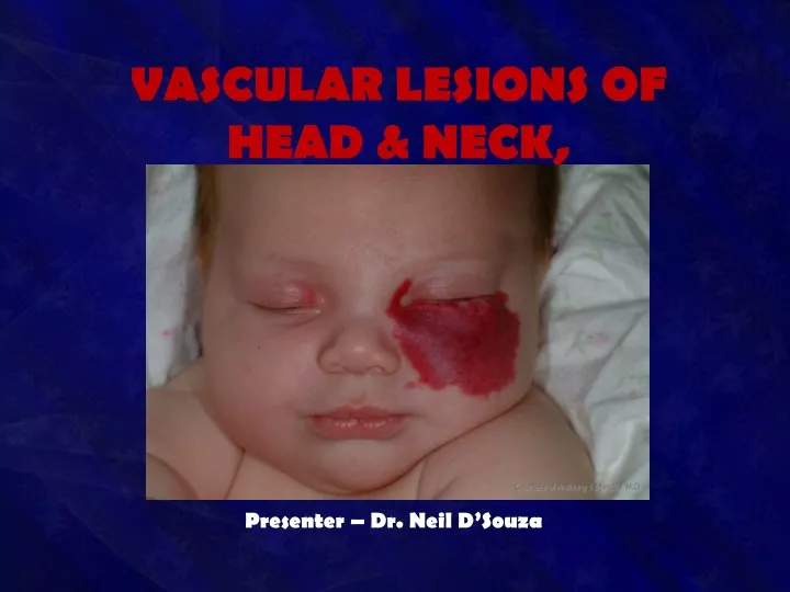
VASCULAR LESIONS OF HEAD & NECK, BED SORES Presenter – Dr. Neil D’Souza
Most common vascular tumour in children (in 10% of term deliveries) • Benign vascular endothelial tumour • Common in girls (3 : 1)
Onset is few weeks after birth • Biphasic growth showing initial rapid growth with gradual involution over 5-7 years • Shows cellular endothelial hyperplasia with increased mast cells
Commonly seen in skin and subcutaneous tissue • Common in head and neck region (60%) • Can occur anywhere in the body like in liver, brain, lungs, other organs
It grows rapidly in first year and 70% involutes in 7 years • Early proliferative lesion is bright red, irregular; deep lesion is bluish coloured • Involution causes colour fading, softness, shrinkage leaving crepe paper like area
Head and neck haemangioma is associated with ocular and intracranial anomalies • Ulceration, bleeding, airway block and visual disturbances are common complications. • Platelet trapping and severe thrombocytopenia • Rare, but important life-threatening complication • Presents as ecchymosis, petechiae, intracranial haemorrhage, massive GI bleed
Raised angiogenic (fibroblastic) growth factor which is secreted in patient’s urine is useful lab investigation
Classification • Capillary • Salmon patch (stork bite) • Strawberry haemangioma • Port-wine stain (naevus flammeus) • Cavernous
Presents at birth • Commonly occurs in the nape of the neck (50%), face, scalp and limbs • Usually involves a wide area of skin
Caused by an area of persistent fetal dermal circulation • With age, it undergoes spontaneous regression and disappears completely (usually in one year) • Hence masterly inactivity is the treatment
May start at birth • If the child is normal at birth; between one to three weeks it may appear as a red mark which rapidly increases in size within 3 months to form strawberry/raspberry haemangioma
Contains immature vasoformative tissues • There will eventually be intravascular thrombosis, fibrosis and mast cell infiltration • It is a true capillary haemangioma
It is 20 times more common than port wine stain. • It is common in white girls • Male to female ratio is 1 : 3 • It is common in head and neck region.
It is clinically compressible, warm with bluish surface. • Bleeding, ulceration can occur after minor trauma and also • It involves skin, subcutaneous tissues and often muscles
After one year of age, it slowly begins to fade, and disappears completelyin 7-8 years (70% in 7 years) • It is the most common haemangioma
It presents at birth and persists throughout life without change • Spontaneous regression will not occur • Due to defect in maturation of sympathetic innervation of skin causing localised vasodilatation of intradermal capillaries
Presents as • Smooth, flat, reddish blue/intensely purple area • Eventually the surface becomes nodular and keratotic • Common in head, neck and face; • Often within the maxillary and mandibular dermatomes of 5th cranial nerve
It is actually a capillary malformation even though considered under haemangioma • It is often associated with • Sturge-Weber syndrome, • Klippel-Trenaunay-Weber syndrome and • Proteus syndrome
Treatment • Wait and watch policy commonly - allowed for spontaneous regression • Pulsed dye laser (diode laser) • Surgical excision and reconstruction • Sclerotherapy/ cryotherapy/ CO2 snow therapy cause unpleasant scarring
Indications for surgery or intervention • Uncontrolled growth • Functional impairment like vision or hearing • Accidental haemorrhage
Feeding vessels may need to be ligated after wide exposure before achieving complete extirpation • Preoperative embolisationfacilitates surgical excision and reduces the operative blood loss
10-year-male with left cheek hemangioma treated with percutaneous glue embolization followed by surgical resection
Once embolisation is done, surgery should be done as early as possible otherwise recurrence occurs with enlarged collaterals • Materials used are—foam, plastic spheres, stainless/platinum steel coils, ethanol, polyvinyl alcohol foam of 1000 μmeters size, and rapidly polymerizing acrylic • Done with interventional radiology underC-Arm guidance
Rapidly growing haemangioma may need systemic/oral and intralesional steroid therapy • Antiangiogenicinterferon 2a may be useful • Life-threatening platelet trapping may be controlled by cyclophosphamide chemotherapy • Haemangioma with drug resistane can be treated with radiotherapy
Presents at birth • Gradually increases in size • Consists of a multiple venous channels • Often mixed with lymphatic component also
Sites: • Head & neck • Limbs, • Tongue, • Liver and other internal organs
Large or multiple cavernous haemangiomas can cause congestive heart failure (hyperdynamic) due to shunting of large quantity of blood • Cavernous haemangioma with dyschondroplasia is called as Maffucci syndrome
Clinical Features • Non-pulsatile swelling • Smooth • Soft • Well localised • Warm • Fluctuant • Compressible • Bluish surface • Non-transilluminant
Usually nontender unless it gets infected or undergoes thrombosis or causes haemorrhage
Differential Diagnosis • Lymphangioma: it is brilliantly transilluminant unless it is infected or fibrosed • Lipoma • Cold abscess • Lymph cyst
Complications • Haemorrhage • DIC • Thrombosis • Infection and septicaemia • Ulceration • Erosion into the adjacent bone • High output cardiac failure
Investigations • Ultrasound with doppler • Angiogram to find out feeding vessel • Platelet count • MRI/MR angiogram to see feeding vessels and deeper extension
Treatment • Sclerosant therapy: • Initial first line of therapy • Causes aseptic thrombosis and fibrosis • Sodium tetradecyl sulphate/ hypertonic saline are used • Often multiple injections are needed to achieve complete required effect • Later excision of the lesion is done