Download
1 / 34
380 likes | 1.47k Vues
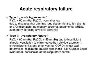
ACUTE RESPIRATORY INFE CTIONS IN CHILDREN. Dr. NITHU ANN Department Of Pediatrics. Introduction. Upper and lower respiratory tract separated at base of epiglottis .

E N D
ACUTE RESPIRATORY INFECTIONSIN CHILDREN Dr. NITHU ANN Department Of Pediatrics
Introduction • Upper and lower respiratory tract separated at base of epiglottis. • Upper respiratory tract consists of airways from the nostrils to the vocal cords in the larynx, including the paranasal sinuses and the middle ear. • The lower respiratory tract covers the continuation of the airways from the trachea and bronchi to the bronchioles and the alveoli.
Introduction • ARI responsible for 20% of childhood (< 5 years) deaths90% from pneumonia • ARI mortality highest in children • HIV-infected • Under 2 year of age • Malnourished • Weaned early • Poorly educated parents • Difficult access to healthcare • Out- patient visits 20-60% • Admissions 12-45%
Factors influencing the incidence of respiratory tractinfections • Poor nutritional status • Poor socio-economic status • Parental smoking • Parasitic infection • Structural abnormalities • Breastfeeding and early weaning • Immunization • HIV incidence • Rainy and cold weather
UPPER RESPIRATORY TRACT INFECTIONS • RHINITIS (COMMON COLD OR CORYZA) • RHINOVIRUSES, ENTEROVIRUSES, CORONAVIRUSES • Acute Tonsillopharyngitis • EAR INFECTIONS (ACUTE OTITIS MEDIA) • VIRUSES,PNEUMOCOCCUS,H.INFLUENZA,MORAXELLA CATARRHALIS • Sinusitis
RHINITIS (COMMON COLD OR CORYZA) • Cause: • Adenoviruses, Influenza, Rhinovirus, Paranfluenza virus, RSV etc. • Rhinitis can also be due to allergy. • Spreads by droplet infection. • Clinical features : • Fever, serous discharge, irritability. • Cervical lymphadenopathy, Nasopharyngeal congestion causing nasal block & difficulty breathing. • Eustachian tube block- serous otitis media
RHINITIS (COMMON COLD OR CORYZA) • Treatment • Relieve Nasal congestion- use saline drops ,avoid decongestants, antihistaminicsare avoided in <6m age. • Nonsedating agents (cetirizine) may be used in allergic rhinitis. • Antipyretics for fever. • Antibiotics are used for secondary bacterial infection only if secretion are purulent, high fever or developing bronchopneumonia.
Acute Tonsillopharyngitis • Viral or bacterial infection of throat causing inflammation of the pharynx & tonsils. • Causative agents: Viral – Adenovirus, Influenza, Parainfluenza, Enterovirus, EBV etc Other – Streptocooci, Mycoplasma pneumonia, Candida • Clinical Features: Fever, Headache, Nausea, Sore throat Refusal to feed in younger children Pharyngo-tonsillar Congestion/Exudates.
Acute Tonsillopharyngitis • Management • Medical -- Rest, Liquid/soft diet, warm saline gargles, cool moist environment. -- Antipyretics, Antibiotics (if bacterial) • Surgical --Tonsillectomy in chronic tonsillitis is controversial. -- Advised for >5 to 6 episodes of tonsillitis/yror tonsillar/peritonsillar abscess.
Acute Lower Respiratory Tract Infections • Epiglottitis • Laryngitis and Laryngotracheobronchitis (LTB) • Pneumonia • Bronchiolitis
Acute epiglottitis • Infection of the epiglottis • Peak incidence :- 1 – 6 years • Male affected more • Bacterial Infection ( H. Influenzae type B ) • Concomitant bacteremia, pneumonia, otitis media, arthritis and other invasive infections caused by H. Influenzae type B.
Acute epiglottitis • CLINICAL FEATURES • High fever, sore throat, dyspnea, rapidly progressing respiratory obstruction • Patient may become toxic, difficult swallowing, laboured breathing, drooling, hyperextendedneck • Tripod position (sitting upright and leaning forward) • Cyanosis , coma • Stridor is a late finding
Acute epiglottitis • EXAMINATION ASSESSMENT OF SEVERITY • DEGREE OF STRIDOR • RESP RATE • H.R • LEVEL OF CONSCIOUSNESS • PULSE OXIMETRY
Acute epiglottitis • DIAGNOSIS: • “Cherry red”appearance of epiglottis on laryngoscopy • Thumb sign on lateral neck radiograph
Acute epiglottitis: Treatment • Need to be managed in ICU, endotracheal intubation may be needed. • Fluid and electrolyte support • I.V AMPLICILLIN100 mg/kg/day OR CEFTRIAXONE 100 mg/kg/day • OTHER OPTIONS • CEFUROXIME OR CEFOTAXIME • CHOLRAMPHENICOL • Treatment Duration :-7-10 DAYS • Rifampicin prophylaxis to close contacts
Acute LTB (Viral croup) • Viral infection leading to mucosal inflammation of the glottic and subglotticregions • Commonly due to influenza(type a), parainfluenza (1, 2, 3) and RSV. • Age :- 6 months – 6 years
Acute LTB (Viral croup) • CLINICAL FEATURES • Initial :- rhinorrhea, mild cough, fever • Later (24-48 hours) :- • Barking cough • Hoarseness of voice • Noisy breathing (mainly on inspiration) • Symptoms worsen at night and on lying down • Children prefer to be held upright or sit in bed.
Acute LTB (Viral croup) • CLINICAL EXAMINATION • Hoarse voice • Normal to moderately inflammed pharynx • Slightly increased resp rate with prolonged inspiration and inspiratory stridor • DIAGNOSIS • Mainly a clinical diagnosis • Radiograph neck :- steeple sign (unreliable)
Acute LTB (Viral croup) [Steeple sign]
Acute LTB (Viral croup) • TREATMENT • Moist or humidified air • Steroids • Reduce the severity and duration / need for endotrachealintubation • Dexamethasone/Prednisolone • Nebulization with budesonide • Nebulization with adrenaline (epinephrine)
Pneumonia • ClassifiedAnatomically as : Lobar or lobularpneumonia, bronchopneumonia, Interstitial pneumonia. • Etiology: • Viral- RSV, Influenza, Parainfluenza, Adenovirus • Bacterial- 0 - 2m : Klebsiella, E.coli, Pneumococci, Staph. 3m-3yr : Pneumococci, H.influenza, Staph. >3yr : Pneumocooci& Staph. • Atypical organisms- Chlamydia, Mycoplasma, Pneumocystis jiroveci, histoplasmosis, coccidioidomycosis. • Others-Ascaris, Aspiration ( food, kerosene, oily nose drops etc).
Danger Signs • High risk of death from respiratory illness – Younger than 2 months – Decreased level of consciousness – Stridor when calm – Severe malnutrition – Associated symptomatic HIV/AIDS
Pneumonia: Radiology Bacterial – Poorly demarcated alveolar opacities with air bronchograms – Lobar or segmental opacification
Pneumonia: Radiology Viral – Perihilar streaking, interstitial changes, -- Air trapping
Pneumonia : Radiology Clues to other specific organisms- • Staphylococcus – areas of break-down • Klebsiella, anaerobes, H. influenza or TB –cavitating or expansilepneumonia • TB, S. aureus, H. influenza -pleural effusion and empyema
Pneumonia: Complications • Empyema • Lung abscess • Pneumothorax • Pneumatocele • Pleural effusion • Delayed resolution • Respiratory failure • Metastatic septic lesions • Meningitis • Otitis media • Sinusitis • Speticemia
Pneumonia : Treatment • Maintain Airway • Oxygen • Hydration • Temperature control • Chest drain :- for fluid or pus collection in chest (empyema) • Antibiotics
Bronchiolitis • Common serious acute lower respiratory infection in infants. • Caused by RSV predominantly; other organisms are influenza, parainfluenza, adenovirus, mycoplasma. • Age - 1 to 6m ( can occur upto 2yrs )
Bronchiolitis • Pathogenesis- • Inflammation of bronchiolar mucosa, edema, mucous plugging. • Bronchiolar narrowing- increased airway resistance. • Air trapping and hyperinflation • Reduced ventilation, CO2 retention, Resp acidosis, Hypoxemia.
Bronchiolitis • Clinical features- • Few days following URTI child presents with tachypnea and respiratory distress. • Dyspnea and Cyanosis may appear. • Prolonged expiratory phase with crepts and rhonchiB/L. • Hyperinflation : liver & spleen may be pushed down. • X-ray : Hyperinflation & Infiltrates.
Bronchiolitis : Treatment • Mild cases can be cared for at home. • Nursed with head & neck elevated to 300 to 400 • Humidified O2 for hypoxemia, keep O2sat >92%. • Maintain Hydration : I.V Fluids. • Nebulization : Adrenaline, 3% NS, Bronchodilators have been tried but efficacy not established. • CPAP, Assisted ventilation may be needed. • Antibiotics have no role.