Download
1 / 43
450 likes | 672 Vues
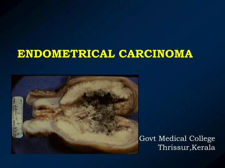
ENDOMETRICAL CARCINOMA. Govt Medical College Thrissur,Kerala. IMPORTANCE???. Recently emerged as the more frequently encountered gynecological cancer accounting for 20-25% of all genital cancers in developed countries.

E N D
ENDOMETRICAL CARCINOMA Govt Medical College Thrissur,Kerala
Recently emerged as the more frequently encountered gynecological cancer accounting for 20-25% of all genital cancers in developed countries. In India incidence is low as 5-7% of all genital cancers,continues to be the second most common of all genital cancers. Incidence of Ca Endometrium : Cervix Western countries India 1:1 1 :8
Recent years there has been an increase in incidence of Ca endometrium • Prolonged life expectancy • Injudicious use of oestrogen • High detection rate • Increasing awareness and screening
INCIDENCE • Peak incidence is 55-70 yrs Perimenopausal 20-25% Women < 45 yrs 5% • Women are either nulliparous or of low parity • Early menarche & late menopause is characteristic • 75% are localised in the uterus when diagnosed • Poorly differentiated in post menopausal and well differentiated and curable in young women
Any factor that increases the exposure of endometrium to unopposed or high osetrogen level, both endogenous and exogenous , increases the risk . • Also linked to the dose and duration of exposure and the risk persists for 10 yrs after hormone exposure.
Oestrogens • Persistant stimulation of endometrium with unopposed oestrogen • (a) Endogenous – PCOD, fibroid uterus, Granulosa cell tumor • (b) Exogenous – OC pills, HRT • Age 75% occur – in > 60 yrs of age • 3. 10 % of post menopausal women are having Ca endometrium
4.Nulliparity and with less children • (30% case) • 5. Late menopause • ↑ incidence if menses fails to stop after 52 years • Obesity • low levels of sex hormone binding globulin levels and this allows free oestrogen to circulate in the body • peripheral conversion of oestrogen to oestrone
7. DUB Chronic non ovulatory cycles 8. Family history Ca endometrium due to genetic predisposition (12-28%) 9. Diet rich in fat → HTN – DM →Ca 10. Tamoxifen antioestrogenic agent with mild oetrogenic activity Prolonged use ↑ incidence
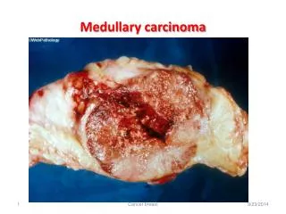
Can be localized or diffuse • Appear as nodule,a polyp,or as a diffuse lesion involving entire uterine cavity. • Extends to endocervix in advanced stage and invades myometrium to varying degree.
Later involves vault by direct spread or suburethral metastasis occurs through retrograde lymphatic or vascular channel • Spreads to adnexa,ovaries as well as to pelvic and para-aortic nodes • Fundal growth spreads to para-aortic lymph nodes via ovarian lymphatics and also to superficial inguinal lymph nodes via round ligament
Microscope • 1. Adeno Ca (Endometrioid Ca) -80% • 2. Adeno squamous Ca • 3. Papillary Ca • 4. Clear cell adeno Ca • 5. Mucinous adeno Ca • 6. Primary squamous cell Ca • 7. Mixed variety
GRADING Grading is based on differentiation, glandular architecture & anaplasia of the cells
Clinical Features • 7-10% are Asymptomatic • Post menopausal bleeding – 75% cases • Premenopausal – menorrhagia or irregular periods • h/o PCOD / HRT may be elicited • HTN ,obese,DM • Clinical features of a bulky uterus(due to growth itself,or due to fibroid or pyometra) may not always be present • In advanced cases cervix is bulky and os patulous with growth protruding through the os,a metastatic vaginal growth is visible near urethra
INVESTIGATIONS PAP SMEAR Only 50% sensitive and not reliable Endometrial cells reveal large round cells with dark nuclei filling most of the cells. ASPIRATION CYTOLOGY Effective in screening high risk Done with Pipelle curette, Issac aspirator,Vibra aspirator, and Novak curette
FRACTIONAL CURETTAGE Histological study of endocervical scraping before dilating the cervix , followed by cervical dilatation and curettage from the isthmus, body and fundus of the uterus separately, so that extent can be evaluated HYSTEROSCOPY Direct visualisation and biopsy Chance of spill of cancer cells into peritoneal cavity limits its use
ULTRASOUND Studying endometrial thickness , detecting polyp and associated ovarian tumour or ovarian metastasis Extension to cervix can also be recognised Post menopausal women normal endometium should not exceed 4 mm in thickness and 10 mm in perimenopausal women DOPPLER ULTRASOUND Revealing a low resistance index of 0.37+/- 0.7 or below is seen in endometrial malignant lesions
SONOSALPINGOGRAPHY Detection of endometrial polyp which could be malignant CA-125 • 35 IU/ml significant • Not specific for CA endometium CT Study extent of spread of lesion localises myometrial infiltration But pelvic nodes < 1cm cannot be identified Superior to MRI in detecting ascites, bowel and omental metastasis
MRI Superior to CT in detection of myometrial involvement and nodal enlargement with 90% detection rate without radiation hazards X-ray Lungs & bone PET-CT Reveals metabolic activity in tissues and is gold standard for staging
DIFFERENTIAL DIAGNOSIS • DUB • Senile vaginitis • Vulval growth • Vaginal growth • Cervical lesions • Uterine tumour • TB endometritis • Atrophic endometritis • Atypical hyperplasia and polypi
STAGING A staging laparotomy is recommended through a midline lower abdominal incision,collect any peritoneal ascitic fluid or washings for cytology ,complete abdominal exploration followed by total abdominal hysterectomy along with bilateral salpingo-oophorectomy and pelvic and para-aortic lymph node sampling.
Surgical staging (FIGO) Stage I – Cancer confined to corpus uteri Stage I a (G123) – Tumor limited to endometrium Stage I b(G123) - Invasion <1/2 myometrium Stage 1 c(G123)- Invasion >1/2 myometrium Stage II – Tumour involves cervix but doesnot extend beyond uterus Stage II a (G123)-Endocervical glandular involvement Stage IIb(G123)- Cervical stromal involvement Stage III- Local and/or regional spread Stage III a (G123)-Tumor invades serosa or adnexa or +ve peritonial cytology Stage III b(G123)-Vaginal metastasis Stage III c(G123)- Pelvic metastasis or paraaortic LN Stage IV- Tumour widespread Stage Iv a(G123)-Tumor involve bladder or bowel mucosa Stage Iv b (G123)-Distant metastasis intra abdominal or inguinal LN
MANAGEMENT Pre Op evaluation 1. Surgery Blood, Urea, X-ray 2. Radiotherapy ECG, LFT, RFT, 3. 3.Combined USG, MRI, Ca 125 4. Chemotherapy Surgery – Is the mainstay Surgical staging,TAH+ BSO + pelvic as well al para aortic lymph node sampling remains the cornerstone in the Mx of early endometrial cancers
Abdomen is opened by a vertical incision that allows a thorough intra-abdominal exploration • Peritoneal washings are obtained from sub diaphragmatic area, paracolic gutters and the pelvis and sent for cytology • After hysterectomy cut open uterus and assess myometrial involvement and cervical extension,tumors size • Tumor differentiationby frozen section • Lymph node sampling or lymphadenectomy is dictated by preoperative grading of tumour,histopathology report and myometrial invasion
Stage 0 – Simple endometrial Hyperplasia – 10-20% Atypical hyperplasia – 70% Elderly TAH + BSO (with removal of ovaries) Prolonged Progesterone therapy Young Patient Progesterone therapy (30-40mg medroxyprogesterone acetate for 6-12 months Life long follow up Levonorgestrel containing IUCD
Stage I – TAH + BSO Pertoneal washing Omental Biopsy, Pelvic or Para aortic LN sampling followed by irradiation after 4-6 wks after surgery No myometrial invasion observation only Myometrial invasion <1/2 thickness. Vaginal vault radiation 6000-7000CGY using colpostats Myometrial thickness >1/2 Whole pelvis ext.irradiation with 6000 CGy over 5-6weeks
L.N. metastasis – whole abdomen irradiation when Pelvic, Common iliac, Para aortic node are involved protecting the liver and kidneys Intracavitary radiation followed by extended hysterectomy after 48 hours in cases of -Highly Anaplastic tumor -Uterine cavity considerably enlarged -Growth extending to cervix -Deeply invasive cancer
Vaginal Hysterectomy- If stage I, well differentiated tumor obese with vaginal prolapse ,should also be combined with laproscopic lymphadenectomy or postoperative radiotherapy
Stage II • Pre-op intracavitary radiotherapy followed by TAH +BSO +Pelvic node dissection • (OR) Wertheims Hysterectomy followed by ext.radiotherapy, • if LN involved dose of 5000CGy over 5 weeks.
STAGES III & IV Inoperable Treated by brachytherapy followed by external radiation Dose – 6000CGy brachytherapy to a depth of 1-1.5cm beneath endometrium and 7000 CGy to vaginal surface with colpostats over 48-72 hours Adjuvant chemotherapy and progestogen therapy prolong remission and improve quality of life.
Progesterons – Excellent response with well differentiated Ca with adequate oestrogen and progesterone receptors 17-alpha Progesterone 1 gm IM weekly MdPA 1gm 1/m weekly or 200mg orally daily Tamoxifen 10 mg twice daily –reduces oestrogen rcps Treatment given for 3 months. If responds give for longer period in reduced dosage. Chemotherapy Doxorubicin 60mg/m2 With cisplatin and paclitaxel
Follow up After initial therapy Every 4 months x 2 years 6 months x 2 years then annually
RECURRENT GROWTHS Occurs within 2 yrs in 50% 3 yrs in 75% Metastasis occurs in vaginal vault , lateral pelvic walls lymph nodes, lungs, liver, brain and bones Distal metastasis occurs in those undergone surgery and post op pelvic radiotherapy
Prognosis Bad Poorly differentiated Ca > Myometrial invasion Advanced stage Advanced age Aneuploid tumors Non endometrioid tumors
5 year survival • Stage 1 - 82% • Stage II -65% • Stage III -44% • Stage IV -15%