Download
1 / 11
110 likes | 249 Vues
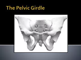
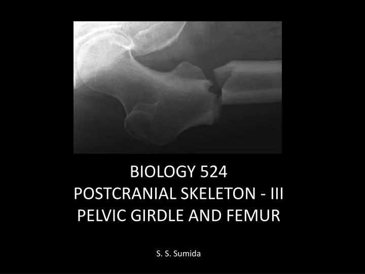
BIOLOGY 524 POSTCRANIAL SKELETON - III PELVIC GIRDLE AND FEMUR S. S. Sumida. The tetrapod pelvic girdle is a tripartite element through it’s history.

E N D
BIOLOGY 524 POSTCRANIAL SKELETON - III PELVIC GIRDLE AND FEMUR S. S. Sumida
The tetrapod pelvic girdle is a tripartite element through it’s history. • Primitively, the regions are not dlearly distinct, but soon after tetrapods are established, a dorsal ILIUM, cranial ventral PUBIS, and caudal ventral ISCHIUM are present. • Internally (medially) the Ilium receives the hypertrophied sacral rib(s). • Laterally, the hip socket for reception of the femur (ACETABULUM) is at the intersection of the three elements.
MORE DERIVED TETRAPODS NOTE particularly in dinosaurs, there are two fundamental hip types, which distinguish the two great groups of dinosaurs: the SAURISCHIAN condition found in Sauropods and Theropods, and the ORNITHISCHIAN condition found in the other great group dominated by herbivorous dinosaurs.
SYANPSIDA • In the synapsid lineage leading to mammals, there is: • A progressive decrease in the size of the ilium • Beginning with therapsids, the development of a larger opening for passage of the obturator nerve, the obturator foramen. It is covered in part by the obturatormembrane that is strong enough to allow muscular attachment. • Basal mammals through primitive placentals have an additional hip element, the EPIPUBIC BONES. They act as a kinetic link between ventral body wall muscles and appendicular muscles.
FEMUR • Like the humerus, the femur is a monobasal articulation primitively. • As with the humerus, primitively it does not have a distinctly defined shaft. • The proximal articular surface is not as complex as that of the humerus. • The ventral surface of the shaft is dominated by an oblique ADDUCTOR RIDGE, which is marked proximally by the FOURTH TROCHANTER, for attachment of aductors and the caudofemoralis muscles. • The adductor ridge is very prominantin diadectomorphs, and some larger basal reptiles.
One of the most well documented changes in the appendicular skeleton is the transition from non-avian theropod dinosaurs to birds. Notably, the femur becomes more flexed at the hip. Note also that the center of mass remains centered approximately over the knee jont.
MAMMALS • As with the humerus, the shaft become more clearly defined. • The spherical head is generally more than 50% of a spherical surface. • The articular head is oriented to allow para sagittal orientation of the hind limb. • The greater trochanter is well developed for attachment of gluteal musculature.