Download
1 / 28
280 likes | 651 Vues
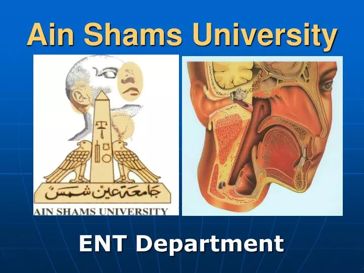
Ain Shams University. ENT Department. The Ear. Trauma to External Ear Haematoma Auris. It is collection of blood under auricular perichondrium.

E N D
Ain Shams University ENT Department
Trauma to External EarHaematoma Auris • It is collection of blood under auricular perichondrium. • It is either due to direct trauma (boxers ear) or spontaneous due to haemorrhagic blood disease or degenerative vascular disease.
Treatment of Haematoma Auris • Aspiration if blood still fluid or • Incision and evacuation if clotted and pressure bandage & prophylactic antibiotics. • If untreated fibrosis of the clot and permanent thickening (cauliflower ear)
Perichondritis Of Auricular Cartilage It is infection of the auricular cartilage which may lead to necrosis. Shrinkage and deformity of the auricle may result. Causes 1. Complication of Haematoma Auris. 2. Complication of surgical operations for chronic otitis media, or of traumatic lacerations of the auricle. 3. Furuncle of the external auditory canal.
Treatment of Perichondritis 1- Antibiotics 2- Wide incision and drainage with excision of the necrotic cartilage.
Traumatic perforation of Tympanic MembraneCauses: 1- Direct trauma • Self inflected. • F.B. in ext. auditory canal or unskilled attempts to remove. • Fractured base of skull extending to attachment of the D.M.
Traumatic perforation of Tympanic Membrane(cont.) 2-Sudden air compression • Hand slap (the most common cause). • Explosions (blast) • Otitic barotrauma. • Over inflation of the Eustachian tube.
Traumatic perforation of Tympanic Membrane(cont.) 3- Sudden water compression - Unskilled ear wash. - Diving or water polo. Symptoms 1- Pain & vertigo may occur at the time of rupture. 2- Deafness, tinnitus & autophony. 3- Air may come out of the ear on nose blowing.
Traumatic perforation of Tympanic Membrane(cont.) Signs: the perforation is in membrana tensa, with irregular or triangular shape and with few blood clots around it. Differential Diagnosis: 1- self inflected There is hesitation marks on the skin of the deep meatus , it is usually in the postro-inferior quadrant of the D.M. and small in size. 2- Pathological perforation
Treatment: 1- Instruct the patient to avoidwater from entering the ear (put cotton with vaseline during bathing)also avoid any ear drops. 2- Avoid blowing of the nose violently. 3- Prophylactic Antibiotics. 4- Myringoplastyoperation: The perforation usually heals within 3 weeks , but there is hope for 3-6 menthes, but if still present, do myringoplasty operation.
Otitic Barotrauma It occurs during descent by aircraft or during diving. Cause : air pressure in high altitude is low and when the aircraft descent rapidly the pressure increase and become more than that in the middle ear so air must go through Eustachian tube to middle ear.
Otitic Barotrauma (cont.) If Eustachian tube does not open (due to edema in its wall as in rhinitis or allergy or during sleep or no swallowing). There will be negative pressure in the middle ear.
Otitic Barotrauma (cont.) • The negative pressure in the middle ear will lead to: • 1- Retractionof the drum membrane that may lead to rupture. • 2- Congestion and edema of m.m. of middle ear followed by transudation of Fluid.
Otitic Barotrauma (cont.) Clinical Picture: 1.Pain and deafness & tinnitus 2.Drum is retracted or even perforated. 3.Drum may be intact and show fluid level behind it (hair lines).
Treatment of Barotrauma Prophylactic treatment: 1) Avoidance of flying with URTI 2) During descent chewing gum, always swallow, do Valsalva, do not sleep “ET are not opened by swallowing during sleep”.
Treatment of Barotrauma (cont.) Curative Treatment: 1- Nasal drops & Systemic nasal decongestant 2- Antibiotics. 3- Inflation of Eustachian tube “ there is 3 methods“ a) Valsalva method. b) Politzer’s method. c) Catheter method (Eustachian Catheterization). 4- If Fluid is present do myringotomy.
Disease of the Middle Ear Congenital Anomalies 1- Middle ear Aplasia (Absent M.E.) it leads to conductive deafness and the only treatment is bone conduction hearing aid. 2- Abnormal Ossicles: as fused , absent , deformed or fixed ossicles. Clinically it lead to conductive deafness and treatment is by ossiculoplasty operation. 3- Other anomalies that maybe found during middle ear surgery. • Dehiscence in the floor and exposure of Jugular bulb. • Dehiscence of the bony Facial Nerve Canal (about 20 %) of the population and other anomalies in the course of facial nerve.
Trauma to the Middle Ear 1- Otitic Barotrauma. 2- Ossicular disconnection. 3- Fracture of temporal bone. 4- F. B. in middle ear
Fractures of Temporal Bone It is type of Fracture base of skull (middle cranial fossa) Types: 1 - Longitudinal Fracture 2 - Transverse Fracture
1)Longitudinal Fracture of Temporal Bone • It is the common type (80%). • Fracture line is in long axis of temporal bone • It involves the tympanic cavity , tympanic membrane and bonyexternal canal. Clinical Picture: - Conductive deafness. - Bleeding through ruptured D.M. and may be CSF otorrhoea. - Facial N. paralysis is uncommon and partial.
2- Transverse Fracture of Temporal Bone • It is the less common type (20%). • Fracture line is at right angle to the long axis of temporal bone. • It involves the labyrinth and or internal auditory meatus. Clinical Picture: - Perceptive deafness (S.N.H.L.). - Vertigo & Nystagmus. - Haemotympanum which may contain also CSF. - Facial N. paralysis is more common and more severe.
Fracture of Temporal Bone(cont.) Investigations: 1- C.T. scan to assess the fracture line. 2- Audiological tests to assess : -Type and degree of deafness. - Stapedial reflex . 3-Tests for Facial nerve function.
Fracture of Temporal Bone Treatment 1- Neurosurgical treatment of the patient (for any associated coma or extradural haemorlrage,….) 2- If CSF Otorrhoea - Prophylactic antibiotics (that cross the blood brain barrier). - Semi sitting position and avoid straining. - Sterile ear dressing. -Neurosurgical repair by fascia late for some cases. 3- Facial Nerve paralysis if incomplete and delayed usually recovers spontaneously but if severe and immediate ( indicating severe nerve injury ) do surgical exploration and nerve suture or graft. 4-Treatment of Traumatic Rupture of D.M. & Ossicles.