Download
1 / 33
330 likes | 406 Vues
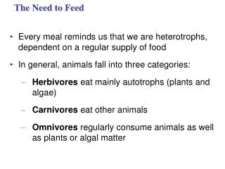
The Need to Feed. Every meal reminds us that we are heterotrophs, dependent on a regular supply of food In general, animals fall into three categories: Herbivores eat mainly autotrophs (plants and algae) Carnivores eat other animals

E N D
The Need to Feed • Every meal reminds us that we are heterotrophs, dependent on a regular supply of food • In general, animals fall into three categories: • Herbivores eat mainly autotrophs (plants and algae) • Carnivores eat other animals • Omnivores regularly consume animals as well as plants or algal matter
An adequate diet must satisfy three needs: • Fuel for all cellular work • Organic raw materials for biosynthesis • Essential nutrients, substances that the animal cannot make for itself
Glucose Regulation as an Example of Homeostasis • Animals store excess calories as glycogen in the liver and muscles and as fat • Glucose is a major fuel for cells • Hormones regulate glucose metabolism • When fewer calories are taken in than are expended, fuel is taken from storage and oxidized
STIMULUS: Blood glucose level rises after eating. Homeostasis: 90 mg glucose/ 100 mL blood STIMULUS: Blood glucose level drops below set point.
Essential Amino Acids • Animals require 20 amino acids and can synthesize about half from molecules in their diet • The remaining amino acids, the essential amino acids, must be obtained from food in preassembled form • A diet that provides insufficient essential amino acids causes malnutrition called protein deficiency
Most plant proteins are incomplete in amino acid makeup • Individuals who eat only plant proteins need to eat a variety to get all essential amino acids • Malnutrition is much more common than undernutrition (HUNGER) in human populations
Essential amino acids for adults Beans and other legumes Methionine Valine Threonine Phenylalanine Leucine Corn (maize) and other grains Isoleucine Tryptophan Lysine
Vitamins and Minerals • Vitamins are organic molecules required in the diet in small amounts • Vitamins are grouped into two categories: fat-soluble and water-soluble • Minerals are simple inorganic nutrients, usually required in small amounts
The main stages of food processing are ingestion, digestion, absorption, and elimination • Ingestion is the act of eating • Digestion is the process of breaking food down into molecules small enough to absorb • Absorption is uptake of nutrients by body cells • Elimination is the passage of undigested material out of the digestive compartment
Small molecules Pieces of food Chemical digestion (enzymatic hydrolysis) Nutrient molecules enter body cells Mechanical digestion Undigested material Food INGESTION ELIMINATION DIGESTION ABSORPTION
In intracellular digestion, food particles are engulfed by endocytosis and digested within food vacuoles • Extracellular digestion is the breakdown of food particles outside of cells • It occurs in compartments that are continuous with the outside of the animal’s body
Mouth Tentacles Gastrovascular cavity Food Animals with simple body plans have a gastrovascular cavity that functions in both digestion and distribution of nutrients Epidermis Mesoglea Gastrodermis Nutritive muscular cells Flagella Gland cells Food vacuoles Mesoglea
More complex animals have a digestive tube with two openings, a mouth and an anus • This digestive tube is called a complete digestive tract or an alimentary canal • It can have specialized regions that carry out digestion and absorption in a stepwise fashion
Crop Gizzard Intestine Esophagus Pharynx Anus Mouth Typhlosole Lumen of intestine (a) Earthworm Foregut Midgut Hindgut Esophagus Rectum Anus Crop Mouth Gastric cecae (b) Grasshopper Stomach Gizzard Intestine Mouth Esophagus Crop Anus (c) Bird
Each organ of the mammalian digestive system has specialized food-processing functions • The mammalian digestive system consists of an alimentary canal and accessory glands that secrete digestive juices through ducts • Mammalian accessory glands are the salivary glands, the pancreas, the liver, and the gallbladder • Food is pushed along by peristalsis, rhythmic contractions of muscles in the wall of the canal • Valves called sphincters regulate the movement of material between compartments
Cardiac orifice Tongue Oral cavity Parotid gland Salivary glands Sublingual gland Pharynx Esophagus Submandibular gland Pyloric sphincter Liver Stomach Ascending portion of large intestine Gall- bladder Pancreas Duodenum of small intestine Ileum of small intestine Small intestine Large intestine Rectum Anus Appendix Cecum
The Oral Cavity, Pharynx, and Esophagus • In the oral cavity, food is lubricated and digestion begins • Teeth chew food into smaller particles that are exposed to salivary amylase, initiating breakdown of glucose polymers • The region we call our throat is the pharynx, a junction that opens to both the esophagus and the windpipe (trachea) • The esophagus conducts food from the pharynx down to the stomach by peristalsis
Bolus of food Tongue Epiglottis up Epiglottis up Pharynx Glottis down and open Esophageal sphincter contracted Glottis Esophageal sphincter relaxed Epiglottis down Esophageal sphincter contracted Larynx Glottis up and closed Trachea Esophagus Relaxed muscles To stomach To lungs Contracted muscles Relaxed muscles Stomach
The Stomach • The stomach stores food and secretes gastric juice, which converts a meal to acid chyme • Gastric juice is made up of hydrochloric acid (pariettal cells) and the enzyme pepsin • Pepsin is secreted as inactive pepsinogen (chief cells); pepsin is activated when mixed with hydrochloric acid in the stomach • Gastric ulcers, lesions in the lining, are caused mainly by the bacterium Helicobacter pylori
Esophagus Cardiac orifice Stomach Pyloric sphincter 5 µm Small intestine Folds of epithelial tissue Interior surface of stomach Epithelium Pepsinogen and HCl are secreted into the lumen of the stomach. Pepsinogen Pepsin (active enzyme) Gastric gland HCl HCl converts pepsinogen to pepsin. Pepsin then activates more pepsinogen, starting a chain reaction. Pepsin begins the chemical digestion of proteins. Mucus cells Chief cells Parietal cells Chief cell Parietal cell
The Small Intestine • The small intestine is the longest section of the alimentary canal (20’-23’) • It is the major organ of digestion and absorption • The first portion of the small intestine is the duodenum, where acid chyme from the stomach mixes with digestive juices from the pancreas, liver, gallbladder, and the small intestine itself • The pancreas produces proteases, protein-digesting enzymes that are activated after entering the duodenum
Bile Liver Gall- bladder Stomach Acid chyme Intestinal juice Pancreatic juice Pancreas Duodenum of small intestine
The liver produces bile, which aids in digestion and absorption of fats • The epithelial lining of the duodenum, called the brush border, produces several digestive enzymes • Enzymatic digestion is completed as peristalsis moves the chyme and digestive juices along the small intestine
Protein digestion Nucleic acid digestion Fat digestion Carbohydrate digestion Oral cavity, pharynx, esophagus Polysaccharides Disaccharides Salivary amylase Smaller polysac- charides, maltose Stomach Proteins Pepsin Small polypeptides Lumen of small intes- tine DNA, RNA Fat globules Polysaccharides Polypeptides Pancreatic amylases Pancreatic nucleases Pancreatic trypsin and chymotrypsin Bile salts Maltose and other disaccharides Fat droplets Nucleotides Smaller polypeptides Pancreatic carboxypeptidase Pancreatic lipase Amino acids Glycerol, fatty acids, glycerides Epithelium of small intestine (brush border) Small peptides Nucleotidases Nucleosides Dipeptidases, carboxy- peptidase, and aminopeptidase Disaccharidases Nucleosidases and phosphatases Nitrogenous bases, sugars, phosphates Amino acids Monosaccharides
Absorption of Nutrients • The small intestine has a huge surface area, due to villi and microvilli that are exposed to the intestinal lumen • The enormous microvillar surface greatly increases the rate of nutrient absorption • Each villus contains a network of blood vessels and a small lymphatic vessel called a lacteal
Key Nutrient absorption Vein carrying blood to hepatic portal vessel Microvilli (brush border) Blood capillaries Epithelial cells Muscle layers Epithelial cells Large circular folds Lacteal Villi Lymph vessel Villi Intestinal wall
Amino acids and sugars pass through the epithelium of the small intestine and enter the bloodstream Capillaries and veins from the lacteals converge in the hepatic portal vein and deliver blood to the liver and then on to the heart After glycerol and fatty acids are absorbed by epithelial cells, they are recombined into fats within these cells These fats are mixed with cholesterol and coated with protein, forming molecules called chylomicrons, which are transported into lacteals
Fat globule Bile salts Fat droplets coated with bile salts Micelles made up of fatty acids, monoglycerides, and bile salts Epithelium of small intestine Lacteal Epithelium of lacteal
The Large Intestine • The large intestine (5’), or colon, is connected to the small intestine • Its major function is to recover water that has entered the alimentary canal • Wastes of the digestive tract, the feces, become more solid as they move through the colon • Feces pass through the rectum and exit via the anus • The colon houses strains of the bacterium Escherichia coli, some of which produce vitamins
Dentition, an animal’s assortment of teeth, is one example of structural variation reflecting diet • Mammals have specialized dentition that best enables them to ingest their usual diet • Herbivores generally have longer alimentary canals than carnivores, reflecting the longer time needed to digest vegetation
Incisors Molars Canines Premolars Carnivore Herbivore Omnivore
Animations and Videos • Bozeman - The Digestive System • Organs of Digestion • Three Phases of Gastric Secretion • Hormones and Gastric Secretion • Hydrochloric Acid Production in Stomach • Balancing Levels of Glucose in the Blood • Blood Sugar Regulation in Diabetics • Reflexes in the Colon
Animations and Videos • Bomb Calorimeter • Calories in a Sandwich • Chapter Quiz Questions – 1 • Chapter Quiz Questions – 2