Download
1 / 63
630 likes | 1.11k Vues
Pakistan Society Of Chemical Pathologists Distance Learning Programme In Chemical Pathology (DLP-2) Lesson No 12 Porphyrias- “Made Easy” By Brig Aamir Ijaz MCPS, FCPS, FRCP (Edin) Professor of Pathology / Consultant Chemical Pathologist AFIP Rawalpindi. Haem in the Body.

E N D
Pakistan Society Of Chemical PathologistsDistance Learning ProgrammeIn Chemical Pathology(DLP-2)Lesson No 12Porphyrias- “Made Easy”By Brig Aamir IjazMCPS, FCPS, FRCP (Edin)Professor of Pathology / Consultant Chemical PathologistAFIP Rawalpindi
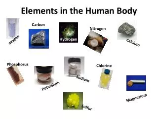
Haem in the Body • Heme is mostly used for its incorporation into haemoglobin and role in red blood cells. • But is also needed for cytochrome P450 function. • Other uses of Haem include: • Catalase • Myoglobin • Nitric oxide synthase
Introduction Porphyrias is a set of diseases that result from enzyme deficiencies in the heme synthesis pathway. Each disease is associated with a deficiency in one of the seven enzymes in the pathway
Free Radicals –The real Culprits • The respective enzymatic deficiencies lead to the accumulation of porphyrins and porphyrin precursors that ultimately produce free radicals. • For acute hepatic porphyrias, delta-aminolaevulinic acid (ALA) and porphobilinogen (PBG) accumulate and produce free radicals via autoxidation. • For erythropoietic porphyrias, uroporphyrins, coproporphyrins, and protoporphyrins accumulate and produce free radicals via the absorption of visible light. • The generated free radicals participate in oxidative stress reactions, such as lipid oxidation and protein crosslinking, that lead to membrane and mitochondrial damage, ultimately promoting cell death. • Loss of negative feedback of heme leads to further accumulation of porphyrins.
Genetics Autosomal Dominant AIP HCP VP EPP Autosomal recessive ADP CEP Acquired PCT
Environmental Factors Due to incomplete penetrance, inheritance of an enzyme deficiency of an autosomal dominant porphyria does not necessarily lead to clinical symptoms. Symptoms of acute porphyrias tend to come in the form of "attacks" which may be induced by other genetic factors or environmental factors, such as agents that promote porphyrin and porphyrin precursor synthesis and/or agents that induce cytochrome P450s.
Q. 1: Porphyria's are usually classified as ‘Acute and Non-acute’, ‘With or Without cutaneous lesions’, pattern of inheritance, ‘congenital or acquired’. Please write name(s) of porphyrias falling in following five categories. Please write only the names of the disorders and NOT the names of effected enzymes (one mark each):a. Acute (neurovisceral) without any cutaneous lesionsb. Acute (neurovisceral) with blistering cutaneous lesionsc. Non-acute (non-neurovisceral) with blistering cutaneous lesionsd. Acute photosensitivity and non-blistering lesionse. Acquired
Classification of Porphyrias a. Acute (neurovisceral) without any cutaneous lesions • Acute intermittent porphyria (AIP) • ALA dehydratase deficiency porphyria (ALADP) • Acute (neurovisceral) with blistering cutaneous lesions • Variegate porphyria (VP) • Hereditary coproporphyria (HCP) • Non-acute (non-neurovisceral) with blistering cutaneous lesions • Porphyria cutanea tarda (PCT) • Hepatic erythropoietic porphyria (HEP) • Congenital erythropoietic porphyria (CEP) • Acute photosensitivity and non-blistering lesions • Erythropoietic porphyria (EPP) • Acquired • Porphyria cutanea tarda (PCT)
Types of Porphyrias Slides courtesy of Dr Shahnaz Noor, QAMC BWP
Types of Porphyrias Cutaneous • Bullous lesions • PCT & HEP • CEP • Non-bullous Lesions • EPP Acute Presentation (Hepatic Porphyrias): • AIP • ADP Mixed Disorders (Acute and Cutaneous manifestations) • VP • HCP
Q. 2: Porphyria's cannot be diagnosed without intelligent use of laboratory investigations. Please answer following questions regarding appropriate use of laboratory tests helpful in various clinical situations (one mark each):a. A 25 years female complains of abdominal pain for the last 24 hours. Her blood counts (CBC) and abdominal ultrasound are normal. She has been referred to you for the exclusion of a porphyria. Name TWO laboratory tests which will be most helpful in reaching the diagnosis of a porphyria and how will you interpret these tests?b. What patient safety issues can arise if a false negative or false positive test result is given in the female patient mentioned in 2 (a) above.C. A 30 year old male has bullous skin lesions which have erupted gradually over a period of one week. His biochemical findings were: ALT: 64 U/L Ferritin: 330 ng/ml HFE gene mutation: Positive Which porphyria comes to your mind in this patient and how will you rule out /diagnose this porphyria by using appropriate laboratory test(s)?
Q. 2: a. A 25 years female complains of abdominal pain for the last 24 hours. Her blood counts (CBC) and abdominal ultrasound are normal. She has been referred to you for the exclusion of a porphyria. Name TWO laboratory tests which will be most helpful in reaching the diagnosis of a porphyria and how will you interpret these tests?Urinary porphobilinogen (PBG) measuremento High levels are suggestive of acute porphyria (AIP, VP, HCP)Urinary δ aminolaevulinic acid (ALA) measurement (and repeat PBG) on same urine sampleo In symptomatic patient with normal PBG, raised ALA levels are seen in ALADP, a very rare form of acute porphyrias and lead toxicity
Q. 2: b. What patient safety issues can arise if a false negative or false positive test result is given in the female patient mentioned in 2 (a) above.Consequences of false positive result• Delay in the life saving treatment appropriate for patient’s actual diagnosis• Analgesic misuse and dependencyConsequences of false negative result • Administration of porphyrinogenic drugs that will aggravate the attack and may increase fatality• Unnecessary surgery e.g. laparotomy for acute abdomen not only have its own risks but also deteriorate the acute porphyria attack
Q. 2: c. A 30 year old male has bullous skin lesions which have erupted gradually over a period of one week. His biochemical findings were:• ALT: 64 U/L• Ferritin: 330 ng/ml• HFE gene mutation: Positive Which porphyria comes to your mind in this patient and how will you rule out /diagnose this porphyria by using appropriate laboratory test(s)?See answers on next slides
Porphyria Cutanea Tarda because of following pointso Nonacute porphyriao Bullous skin lesionso Male predominanceo Raised ALT provides evidence of hepatocellular damageo HFE gene mutation has strong positive association with PCT
Laboratory investigationso Urine porphyrins – markedly increased- Chromatographic separation based on carboxyl number reveals predominance of 8- and 7-carboxyl porphyrin fractions, with lesser amounts of 6-, 5-, and 4-carboxyl porphyrins, reflecting uroporphyrinogen decarboxylase (UROD) defect.o Serum or plasma porphyrins- also show increased polycarboxylated porphyrinso Fecal porphyrins – raised. Fractionation reveals complex pattern with increased isocoproporphyrino Urine ALA and PBG – normal which exclude all acute porphyriaso Erythrocyte porphyrins – within reference range o Plasma fluorescence peaks at 620 nm o UROD enzyme activity in red blood cells- markedly reduced and is diagnostic of PCT
Q. 3: Some tests of porphyrias are essential in any lab for the diagnosis of these disorders. Please answer following questions regarding porphyria related tests (one mark each):a. How do you test Porphobilinogen in urine and what precautions you will adopt in collection of urine sample?b. Write TWO lines about any ONE method used for analysis of porphyrins in plasma.
Q 3a. Porphobilinogen tests in urine Qualitative or semi quantitative method • screening method • formation of red colored PBG-Ehrlich compound on addition of Ehrlich reagent to urine that can be separated by solvent extraction • rapid, cheap and still widely used but has poor sensitivity and specificity • Most important interference: Urobilinogen
Q 3a. Porphobilinogen tests in urine (Cont) Quantitative method • Preferred method for measurement of PBG in urine • Spectrophotometric quantification of the red product formed by PBG reaction with 4-dimethylaminobenzaldehyde in acid (Ehrlich’s reagent) at 550 nm after removal of urobilinogen and other interfering substances by anion exchange chromatography. Results should be expressed as µmol/mmol creatinine. HPLC Tandem mass spectrometry • Sensitive and accurate but time consuming
Q 3a. Porphobilinogen tests in urine (Cont) Specimen collection for urinary porphobilinogen • Best specimen is freshly voided spot urine preferably early morning, 10-20ml without any preservative • Must be collected in colored bottle and kept well protected from light • If any delay in transport or analysis and after analysis, specimen must be kept at 4oC for 48 h and at -20oC for 1 month.
b. Write TWO lines about any ONE method used for analysis of porphyrins in plasma.Fluorescence emission spectroscopy • It utilizes the property of porphyrins to fluoresce at neutral pH between 610-640 nm depending upon their structure and protein complex• Simple method that can differentiate among various cutaneous porphyrias
Q. 4 Heme is an important prosthetic group in many important substances of the body. Which of the following tissues / organs are the most active site of heme synthesis:a. Bone marrowb. Kidneysc. Liverd. Lungse. Muscles a. Bone marrow
Biosynthesis of Heme Slides courtesy of Dr Anwar Magsi, PNS SHIFA Karachi
Biosynthesis of Heme • Synthesized in every human cell • Liver (15%): • Bone Marrow (80%)
Q 5. Two different delta aminolaevulinic acid synthase (ALAS) enzyme systems have been found. Only ONE of the following features is common in ALAS1 and ALAS2 enzymes :a. Down-regulation by heme b. First step in the synthesis of hemec. Liver is the major locationd. Mutations have been described in the genes coding these enzymese. Up-regulation by iron b. First step in the synthesis of heme
Two Types of ALAS ALAS-1 Present in liver and all other tissues for production of haem used for non-haemoglobin substances e.g. Cytochrome P450 ALAS-2 Present in erythrocytes for production of Haemoglobin
Q 6. The most common human porphyria is:a. Acute intermittent porphyria b. ALA dehydratase porphyria c. Congenital erythropoietic porphyria d. Porphyria cutanea tarda e. Hereditary coproporphyria . d. Porphyria cutanea tarda
Two Most Important Disorders • Acute intermittent porphyria (AIP) --- Most common acute • Porphyria Cutanea Tarda (PCT)-------The commonest
Acute intermittent porphyria (AIP) • 2nd most common form of porphyria • Caused by deficiency of PGB deaminase • Metabolite porphobilinogen accumulates in cytoplasm • Symptoms: • Localized abdominal pain • Urinary symptoms • Peripheral neuropathy • raised concentration of urinary porphyrins • Treatment • Haematin, Heme arginate • Do not cure but reduces symptoms • Inhibit ALA synthase which occurs at the beginning of heme biosynthesis
A Supplementary QuestionThe commonest presenting clinical feature of Acute Intermittent Porphyria is:a. Abdominal painb. Dark urinec. Muscular weaknessd. Severe constipatione. Tachycardia Best answer: a. Abdominal pain
Symptoms of Acute Forms • Originate mainly in nervous system • Symptoms last around 1-2 weeks • Possible mechanisms include damage by free radicals, direct neurotoxicity of ALA, and the deficiency in nervous tissue Symptoms: • Severe abdominal pain • Muscle weakness and pain, tingling, or numbness and possibly paralysis • Pain in arms, legs, back • Constipation • Vomiting • Diarrhea • Insomnia • Seizures and Confusion • Anxiety and paranoia • Fever
Symptoms of Cutaneous Forms • Occur most commonly with exposure to sunlight • Mainly skin symptoms that occur • Due to excess porphyrins that accumulate in surface of skin • Symptoms: • Fluid filled blisters • Changes in pigmentation • Breakdown (necrosis) of the skin when exposed to sunlight • Overall skin can become scarred, brown, blotchy and fragile
Q 7: Poisoning of which of the following metals resembles acute porphyria due to accumulation of ALA:a. Arsenicb. Copperc. Irond. Leade. Zinc d. Lead
Lead Toxicity and Porphyrias Lead exposure causes increased ALA due to replacement of Zn by Lead in the enzyme. The condition resembles ALADP Also called ‘plumbporphyria’
Q 8: A 31 year female doctor has recently been diagnosed as a patient of Acute Intermittent Porphyria. She shows you a list of factors she has downloaded from internet to prevent precipitation of an acute attack. The list is given below, which of these factors she has wrongly noted:a. Ethanolb. High carbohydrate dietc. Non-steroidal anti-inflammatory drugsd. Oral contraceptive pills e. Smoking b. High carbohydrate diet
Treatment for Acute Forms Carbohydrate such as glucose To help limit the synthesis of porphyrins Phlebotomy To reduce excessive iron stores which improves heme synthesis Sedatives to help with anxiety Pain medications such as opiates Haem arginine Haem inhibit ALA synthase and the accumulation of toxic precursors
Q 9 : In the 1950s, in eastern Turkey an outbreak of Porphyria Cutanea Tarda (PCT) occurred in thousands of adults and children after consumption of stored wheat to fight famine. This outbreak of PCT was a result of:a. Long exposure to sun during queues for wheatb. Periods of prolonged starvation due to faminec. Poor hygiene of the dwellersd. Treatment of wheat with a fungicide e. Tuberculosis due to malnutrition d. Treatment of wheat with a fungicide
Q 10: PCT has been found to be associated with which of the following viral infections:a. Cytomegalovirusb. Ebola virusc. Epstein Barr virusd. Hepatitis C viruse. Human papilloma virus d. Hepatitis C virus
Q 11: An 18 months infant has been referred to you for the investigation of porphyria. His mother describes that whenever she takes his son in the sun, his face and back of hands immediately get red and the child starts crying with pain. On examination you find no lesions on the face or hands, but his Serum AST levels are 2 folds increased. His laboratory investigations related to porphyrias carried out in your lab shows following results: • Urine Porphyrin 25.0 nmol/mmol creat (0 - 35 ) • Urine PBG 0.8 µmol/mmol creat (0 - 1.5 ) • Urine ALA 1.2 µmol /mmol creat (0 - 3.8 ) • Urine Creatinine 4.2 mmol/L• Urinary porphyrin fractions: Not increased • Plasma fluorescence peak at 630 nm • Erythrocyte total protoporphyrins: markedly increased• Erythrocyte metal-free protoporphyria: Markedly increased (92% of total Erythrocytic protoporphyrins) • Erythrocyte Zinc Protoporphyrins: Marginally increased (8% of total Erythrocytic protoporphyrins)• Gene Mutation Studies: Ferrochelatase gene mutation detectedWhat is the most probable diagnosis in this patient? a. Erythropoietic protoporphyriab. Hepatoerythropoietic porphyriac. Lead poisoningd. Porphyria cutanea tarda e. X-linked protoporphyria a. Erythropoietic protoporphyria
Erythropoietic Protoporphyria • Caused by deficiency of Ferrochelatase • Autosomal dominant • Photosensitivity- can be managed by limiting exposure
Q 12: A 2 years Asian boy presented with abdominal pain, vomiting; neuropathic pain and neuropathy. His urine tests showed following results: • Total Porphyrin 64.0 nmol/mmol creat (0 - 35 ) • Porphobilinogen 0.8 µmol/mmol creat (0 - 1.5 ) • Delta-Aminolaevulinic Acid 7.2 µmol /mmol creat (0 - 3.8 ) • Creatinine 3.9 mmol/L • Plasma fluorescence peak at 620 nmWhat is the most probable diagnosis in this patient? a. Acute intermittent porphyriab. Aminolaevulinic acid dehydratase porphyria c. Congenital erythropoietic porphyriad. Erythropoietic protoporphyria e. Hepatoerythropoietic porphyria b. Aminolaevulinic acid dehydratase porphyria
Q 13: A 3 month infant presents with blisters on the skin and photosensitivity. Mother also tells about reddish-brown coloration of the diapers of the infant. He also has hemolytic anaemia and splenomegalyHis laboratory investigations showed following results: • Urine Total Porphyrin 71.0 nmol/mmol creat (0 - 35 ) • Urine PBG 0.68 µmol/mmol creat (0 - 1.5 ) • Urine ALA 1.9 µmol /mmol creat (0 - 3.8 ) • Urine Creatinine 3.1 mmol/L • Plasma fluorescence peak at 618 nm • Urinary porphyrin fractions: o Uroporphyrin I : Increased o Coproporphyrins I: Increased• Erythrocyte total protoporphyrins: markedly increased• Erythrocyte metal-free protoporphyria: Markedly increased (22% of total Erythrocytic protoporphyrins) • Erythrocyte Zinc Protoporphyrins: Marginally increased (68% of total Erythrocytic protoporphyrins)• Gene Mutation Studies: UROS gene mutation detectedWhat is the most probable diagnosis in this patient? a. Congenital erythropoietic porphyriab. Hepatoerythropoietic porphyriac. Hereditary coproporphyria d. Porphyria cutanea tarda e. Variegate porphyria a. Congenital erythropoietic porphyria