Download
1 / 32
390 likes | 618 Vues
The Major Histocompatibility Complex And Antigen Presentation. W. Robert Fleischmann, Ph.D. Department of Urologic Surgery University of Minnesota Medical School rfleisch@umn.edu (612) 626-5034. Objectives. Learn the identity and biological roles of MHC molecules.

E N D
The Major Histocompatibility Complex And Antigen Presentation W. Robert Fleischmann, Ph.D. Department of Urologic Surgery University of Minnesota Medical School rfleisch@umn.edu (612) 626-5034
Objectives • Learn the identity and biological roles of MHC molecules. • Learn the major classes and subclasses of MHC molecules and their roles in immunity. • Understand genetic polymorphism and its significance for MHC molecules. • Understand haplotypes and their significance. • Learn the general structure of Class I and Class II MHC molecules • Learn how and where Class I and Class II MHC molecules bind antigenic peptides.
Pamela Lake, age 9 months, is brought to the clinic for evaluation of possible immunodeficiency. She has a 4-month history of recurrent pneumococcus and Haemophilus infections, despite having been vaccinated for these agents at appropriate times. What tests would you employ to evaluate Pamela and why?
Pamela’s total WBC count and, her differential count were normal. Her RBCs and platelets were normal. Proliferation studies showed normal proliferation following both T cell and B cell mitogen stimulation. However, there was no immune response to antigens, such as poliovirus and tetanus toxoid (for which she had already been vaccinated). A nearly complete absence of antibodies was noted. Only a low level of IgG was detected. Why was only IgG detected? What are your thoughts?
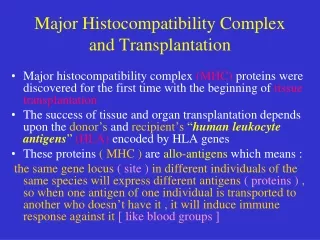
What Are MHC Molecules? • MHC = major histocompatibility antigens • Aka the HLA (human leukocyte antigen) complex in humans and the H-2 complex in mice • They were originally recognized for their involvement in rejection of tissues exchanged between two unrelated organisms. • Now, we know that MHC molecules play an essential role in antigen recognition and presentation to the immune system.
The MHC Region of the Genome Is Large • The MHC gene complex contains more than 100 separate loci (or genes) subdivided into Class I, Class II, and Class III MHC molecules. • Class I MHC molecules present antigen to CD8+ cytotoxic T cells. • Class II MHC molecules present antigen to CD4+ helper T cells. • Other MHC proteins have been grouped together as Class III MHC molecules. They include a diverse group of proteins. • Complement proteins • TNF- and TNF-. • Other loci encode enzymes, heat shock proteins, and some molecules involved in antigen processing.
Importance of MHC Class I and Class II Molecules • B cells express B cell receptor that recognizes antigen without the need for other factors (specific antibody + Ig- and Ig-. • T cells express T cell receptor (or, more accurately, TCR + CD3) that recognizes antigen only when the antigen has been processed and presented to the T cell receptor as an epitope bound to another molecule. This other molecule is an MHC molecule.
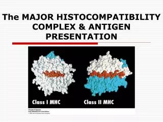
Three Major Classes of MHC Molecules • Class I MHC genes • Present on nearly all nucleated cells • Presents antigen to CD8+ cytotoxic T cells • Class II MHC genes • Present primarily on professional antigen-presenting cells (Ms, dendritic cells, B cells) • Presents antigen to CD4+ helper T cells • Class III MHC genes • Complement proteins • TNF- and TNF- Note: Class I and Class II are structurally similar and bind and present antigen. Class III molecules are very different structurally and do not play a role in antigen presentation.
Class I MHC Have Subclasses • Three “classical” Class I gene loci • MHC Class I gene B encoding HLA-B • MHC Class I gene C encoding HLA-C • MHC Class I gene A encoding HLA-A • There are a number of additional, non-”classical” Class I gene loci • The MHC gene locus D has since been identified to constitute the Class II genes • MHC Class I genes E, F, G, H • Each of the Class I gene products associates with another peptide, 2 microglobulin, when they are expressed on the cell surface.
Class II MHC Have Subclasses • Each Class II MHC gene locus encodes two peptides, an subunit and a subunit that are expressed together on the cell surface. • Three major Class II gene loci • MHC Class II gene DP encoding DP • MHC Class II gene DQ encoding DQ • MHC Class II gene DR encoding DR
Each Major Class of MHC Has Subclasses Kuby Immunology
Each Locus of Class I and Class II MHC Is Highly Polymorphic • There are many alleles for each classical Class I and Class II locus. • This polymorphism • Gives us our unique identity. • Permits recognition of self versus non-self. • Is an impediment to transplantation.
Medical Significance of MHC Polymorphism • This polymorphism • Affects the ability to make an immune response. • Affects the resistance or susceptibility to infectious diseases. • At least part of an individual’s ability to develop an immune response is due to the individual’s MHC genes. • Affects the susceptibility to autoimmune diseases and allergies. • HLA-B27 bearers are 90 times more likely to develop ankylosing spondylitis (destruction of vertebral cartilage). • HLA-DR2 bearers 130 times more likely to develop narcolepsy. • HLA-A3/B14 bearers are 90 times more likely to develop hemochromatosis (too much iron absorption, deposited in and damages internal organs).
Haplotypes • Since Class I and Class II genes are closely linked, recombination within the MHC region is rare. • Thus, we inherit Class I and Class II loci from each of our parents as a cassette of gene alleles. • This cassette is called a haplotype. • Our Class I and Class II make-up is composed of two haplotypes, one maternal and one paternal. • The alleles of each Class I and Class II locus are co-dominantly expressed in the same cells. • For successful transplantation without immunosuppression, a recipient would have to have the same gene alleles within his/her two haplotypes as the donor (syngeneic). • Generally, only identical twins are syngeneic. • Congenic, differ only in one locus.
A Single Cell Expresses MHC Molecules That Represent the Casette of MHC Molecules Kuby Note this example is for the mouse
MHC Class I Polymorphisms • For MHC Class I • 370 A alleles • 660 B alleles • 190 C alleles • A total of 370 x 660 x 190 = 4.6 x 10⁷ Class I haplotypes for A, B, and C. (~2 x 10¹⁵ total) • Class I E, F, G, and H are called Class Ib proteins and are less polymorphic than A, B, and C proteins. Class I E and G proteins bind antigen peptides but are involved in NK cell recognition. • Most of the polymorphism differences are in the cleft region (antigenic peptide binding site).
MHC Class II Polymorphisms • For MHC Class II • DR Locus • 3 DR alleles • 400 DR 1 alleles • ≥74 DR 2-9 alleles • DQ Locus • 28 DQ 1 • 62 DQ 1 • DP Locus • ≥19 DP 1 • 118 DP 1
Stylized Structure of Class I and Class II Molecules 45 kDa chain 12 kDa 2 microglobulin chain 33 kDa chain 28 kDa chain Kuby
Ribbon Diagrams of Class I & II Molecules Resident peptide Resident peptide Roitt
General Features of Peptide Binding Clefts of Class I & II Molecules • Similar structural features to peptide binding clefts of both Class I & II • There are up to 6 different Class I molecules and up to 12 different Class II molecules (mixing and matching of different and subunits for a given set of alleles). • Thus, binding of peptide to the peptide binding clefts cannot be as strong as peptide binding to antibody and T cell receptors.
Activation of T Cell by Antigenic Peptide Bound to MHC • Class I MHC presents antigen to CD8+ cytotoxic T cells. • If there are appropriate co-stimulatory signals and cytokines produced, activation of the cytotoxic T cells may occur. • Activation of cytotoxic T cells results in the death of cells expressing the recognized antigen. • Class II MHC presents antigen to CD4+ helper T cells. • If there are appropriate co-stimulatory signals and cytokines produced, activation of the helper T cells may occur. • Activation of helper T cells may result in the activation of B cells to proliferate and differentiate into plasma cells. • Activation of helper T cells may result in the activation of CD8+ cytotoxic T cells to proliferate and attack antigen bearing cells or in the ability of B cells to become antibody-producing plasma cells.
T Cells Show Self-MHC Restriction • CD4+ and CD8+ T cells from an individual recognize antigenic peptides bound to their own MHC proteins. • They do not recognize antigenic peptide bound to allogenic MHC proteins. Syngeneic MHC Allogeneic MHC Syngeneic MHC Cognate Antigen Cognate Antigen Non-cognate Antigen
Role of Antigen-Presenting Cells • Processing of antigen is required for recognition of an antigen by T cells. • Most cells can present antigen with Class I MHC. • This includes the presentation of foreign antigen and self-antigens. • Recognition of antigen bound to Class I is by CD8+ T cells. • Professional antigen-presenting cell present antigen with Class II MHC. • Recognition of antigen bound to Class II is by CD4+ T cells.
Professional Antigen-Presenting Cells • Dendritic cells are the most effective • Constitutively express high levels of Class II MHC molecules • Constitutively express B7 and other costimulatory molecules • Macrophages • Must be activated by phagocytosis to express Class II molecules • Must be activated to express costimulatory molecules • B cells • Constitutively express class II MHC molecules • Must be activated by antigen binding to antibody before they express costimulatory molecules
Different Antigen-Processing and Presentation Pathways for Different MHC Molecules • Presentation of antigen on Class I molecules requires intracellular protein synthesis of the antigen. • Presentation of antigen on Class II molecules requires the endocytic uptake of antigen.
Pamela Lake Several additional studies are performed on Pamela’s blood cells. Her CD4+ T cells produce IL-2 and IFN- when stimulated with mitogen. Her macrophages phagocytosed agarose beads.
Pamela Lake Using a mixture of polyclonal antibodies to MHC molecules, Pamela’s macrophages and B cells were found to be devoid of HLA Class II antigens. HLA Class I antigens were present on all WBCs. A lack of HLA expression is can be caused by a lack of one of several transcription factors. Probing mRNA from Pamela’s cells indicates that regulatory factor X (RFX) is mutant. RFX is responsible for recognizing the X box promoter sequence in the HLA gene. What can be done for Pamela?
Pamela Lake Pamela is scheduled for a bone marrow transplant.
B Cell Receptor • The B cell receptor is composed of mIg together with Ig- and Ig-. • Membrane-bound IgM and IgD (and IgA, IgE, and IgG) has a very short cytoplasmic tail and cannot transmit a transmembrane signal. • When mIg is bound by its antigen, it associates with the heterodimer Ig- /Ig- to form the B cell receptor complex. • The B cell receptor complex transmits a transmembrane signal through the cytoplasmic tails of Ig- /Ig- when the mIg is bound by its antigen.