Download
1 / 41
410 likes | 519 Vues
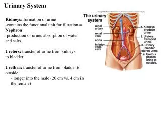
Urinary System. John Minor and Jeremiah Shaw. What are the major parts of the urinary system?. Celiac trunk. Esophagus (cut). Left adrenal gland. Diaphragm. Left kidney. Inferior vena cava. Left renal artery. Left renal vein. Right adrenal gland. Right kidney. Hilum. Superior

E N D
Urinary System John Minor and Jeremiah Shaw
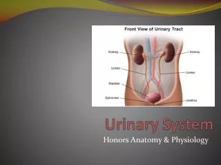
Celiac trunk Esophagus (cut) Left adrenal gland Diaphragm Left kidney Inferior vena cava Left renal artery Left renal vein Right adrenal gland Right kidney Hilum Superior mesenteric artery Aorta Ureter Quadratus lumborum muscle Left common iliac artery Iliacus muscle Gonadal artery and vein Psoas major muscle Rectum (cut) Urinary bladder Anterior view Diagram of Urinary System
Parts of Urinary System • Kidneys – organs that have excretory functions and produce urine • Ureters – tubes leading from the kidneys to the urinary bladder • Urinary Bladder – a muscular sac for temporary storage of urine • Urethra – a tube that conducts urine to the exterior
Functions of Urinary System • Regulation of blood pressure • Regulation of concentrations of ions in plasma • Stabilizing blood pH • Conserving valuable nutrients • Assisting liver in detoxifying poisons
Structure of the Kidney Cortex Renal capsule Medulla Medulla Renal pyramids Renal pyramid Renal sinus Renal sinus Connection to minor calyx Adipose tissue in renal sinus Minor calyx Major calyx Renal pelvis Hilum Renal pelvis Major calyx Hilum Minor calyx Renal lobe Renal papilla Ureter Renal papilla Renal columns Ureter Renal lobe Renal capsule (a) (b)
Anatomy of Kidneys • The kidneys lie on each side of the vertebral column between T12 and L3 • Surrounded by three layers of connective tissue: renal capsule, adipose capsule and the renal fascia • Average kidney: 10cm x 5.5cm x 3cm, weighing 150g • Left kidney lies slightly superior to the right kidney
Anatomy of Kidneys (cont.) • Hilum: entry for the renal artery and nerves and an exit for renal vein and ureter • Renal sinus: internal cavity within the kidney • Renal Pyramids: Triangular structures with bases at the cortex and tips at the renal sinus • Renal Medulla: structure containing 6 - 18 renal pyramids • Renal Columns: bands of tissue that separate renal pyramids
Anatomy of Kidney (cont.) • Cortex – superficial part of kidney • Renal Papilla – the tip of each renal pyramid • Renal Lobe – consists of a renal pyramid, the overlying renal cortex and adjacent columns • Renal Pelvis – a large funnel that drains the kidneys
Afferent arterioles Interlobular vein Interlobular artery Arcuate artery Arcuate vein Interlobular veins Interlobular arteries Renal pyramid Nephron Interlobar arteries Interlobar vein Interlobar artery Segmental artery Suprarenal artery (b) Renal artery Renal vein Renal artery Renal vein Segmental arteries Interlobar arteries Interlobar veins Interlobar veins Arcuate veins Arcuate arteries Arcuate veins Arcuate arteries Interlobular veins Interlobular arteries Venules Afferent arterioles (a) NEPHRONS Glomerulus Peritubular capillaries Efferent arteriole (c) Blood flow of kidney
Blood flow of kidney • The kidney receives blood from the renal artery and blood is removed by the renal vein • Through a system of various types of arteries and veins blood enters and exits the kidney • At the lowest level the blood is passed through the nephron which starts urine production
Proximal convoluted tubule Distal convoluted tubule Reabsorption of water, ions, and all organic nutrients Secretion of ions, acids, drugs, toxins Variable reabsorption of water, sodium ions, and calcium ions (under hormonal control) NEPHRON Renal tubule Capsular space Glomerulus Efferent arteriole COLLECTING SYSTEM Afferent arteriole Bowman’s capsule Descending limb of loop begins Ascending limb of loop ends Collecting duct Variable reabsorption of water and reabsorption or secretion of sodium, potassium, hydrogen, and bicarbonate ions Renal corpuscle Thick ascending limb Production of filtrate Thin descending limb Ascending limb Descending limb Papillary duct KEY Delivery of urine to minor calyx Water Loop of Henle Minor calyx Solutes Filtrate Further reabsorption of water (descending limb) and both sodium and chloride ions (ascending limb) Variable reabsorption or secretion
Anatomy of the nephron • The nephron consist of two parts, the renal corpuscle and the renal tubule • The renal tubule is a long tubular passage way measuring about 50mm • The renal corpuscle is a spherical structure consisting of the Bowman’s capsule and the glomerulus • The renal tubule consist of the proximal convoluted tubule (PCT), distal convoluted tubule (DCT), and the Loop of Henle
Anatomy of the Nephron (cont.) • Glomerulus – system of about 50 intertwining capillaries • Bowman’s Capsule – a chamber that holds the glomerulus with a visceral epithelium
Filtration • The renal corpuscle is the site where filtration occurs • Blood pressure forces water and other solutes out of the glomerular capillaries • This process produces a primarily protein free solution called filtrate
Entry into the renal tubule • After filtrate is formed it enters the renal tubule • As filtrate travels through the renal tubule it becomes tubular fluid with more of the characteristics of urine • The renal tubule has 3 major functions: • Reabsorbing all useful organic nutrients in the filtrate • Reabsorbing more than 90% of water in the filtrate • Secreting any waste products that failed to enter the renal corpuscle through filtration
The Proximal Convoluted Tubule (PCT) • The PCT is the first segment of the renal tubule • The lining of the PCT is a simple cuboidal epithelium whose surface bears microvilli • Reabsorption is the main function of the PCT and it reabsorbs organic nutrients, ions, water and plasma proteins (if present)
Loop of Henle • The second part of the renal tubule is the Loop of Henle • The Loop of Henle is divided into the descending limb and ascending limb • The descending limb flows toward the renal pelvis and is lined with a cuboidal epithelium • The ascending limb flows toward the renal pelvis and is lined with a squamous epithelium
Distal Convoluted Tubule (DCT) • The third part of the renal tubule is the DCT • It is lined with a cuboidal epithelium that is not lined with microvilli • The DCT is necessary for three vital processes • Selective reabsorption of Na and Ca ions • Active secretion of ions, acids, drugs and toxins • Selective reabsorption of water
The Collecting System • The last segment of the nephron is the collecting system • The nephron drains into a collecting duct, which drains into a larger papillary duct, which empties into the minor calyx • Final osmotic pressure and urine volume is determined by the collecting system
General Characteristics of Urine • pH range: 4.5 – 8.0 (average 6.0) • Specific Gravity: 1.003 – 1.030 • Water Content: 93 - 97% • Color : Clear yellow • Odor: Varies with composition
Organic Waste Products • Urea – most abundant organic waste, generate from the breakdown of amino acids (21g) • Creatinine – generated in skeletal muscle through the breakdown of creatine phosphate (1.8g) • Uric Acid – waste product from recycling the nitrogenous base in RNA molecules (.48g)
Basic Processes of Urine Formation • Filtration – blood pressure forces water from the blood across the walls of the glomerular capillaries • Reabsorption – the removal of water and solutes from the filtrate and their movement across the tubular epithelium • Secretion – the movement of solutes from the outside of the tubular epithelium (peritubular fluid) to the inside
Glomerular Filtration Rate (GFR) • GFR is the amount of filtrate produced each minute • On average a GFR is about 125ml a minute, that is around 180 liters of filtrate a day • About 99% of filtrate is then reabsorbed by the renal tubules • One can test GFR with a creatinine clearance test • The GFR can be regulate automatically or with the use of hormones
Middle umbilical ligament Ureter Lateral umbilical ligament Rugae Ureteral openings Center of trigone Neck Internal urethral sphincter Prostate gland External urethral sphincter (in urogenital diaphragm) Prostatic urethra Membranous urethra Urinary Bladder
Anatomy of the urinary bladder • The urinary bladder is a hollow muscular organ that is a temporary reservoir for urine • The superior surface of the bladder is covered with a peritoneum and is stabilized by the middle umbilical ligament and lateral ligaments • The interior surface of the urinary bladder is thrown into folds called rugae • The ureters enter the bladder through ureteral openings and meets the center of trigone • The center of trigone lies superior to the neck which opens to the urethra
Anatomy of urinary bladder (cont.) • Internal Urethral Sphincter – (sphincter vesicae) an involuntary sphincter that allows urine to leave the bladder to the urethra. • External Urethral Sphincter – a circular band of muscles that is under voluntary control to allow for urination
Histology of the urinary bladder • The wall of the urinary bladder has three layers: mucosa, submucosa and muscularis • Muscularis layer has two layers, longitudinal smooth muscular and circular muscle • The two muscle layers form the detrusor muscle, which contracts to expel urine out the urethra
Anatomy of the ureters • The ureters are a pair of muscular tubes that extend from the kidneys to the urinary bladder • They are approximately 12in. in length • The ureters are made from three tissues: - An inner transitional epithelium layer - A middle muscle layer made up of circular and longitudinal layers - An outer connective tissue in conjunction with the renal capsule
Micturition Reflex • The process of urination is coordinated by the micturition reflex • The urge to urinate arises when the urinary bladder contains around 200ml of urine • Nerves receive the signal that the bladder is expanding • The detrusor muscle contracts causing relaxation of the internal and external urethral sphincters
What are the complications that occur with the urinary system?
Urinary issues • Aminoaciduria – amino acid loss in the urine • Calculi – insoluble deposits that form within the urinary tract • Glomerulonephritis – an inflammation of the renal cortex • Hematuria – the presence of blood in urine • Polycystic Kidney Disease – an inherited abnormality that affects the development of the kidney tubule • Proteinuria – the presence of protein in urine • Renal failure – an inability for kidneys to function to maintain homeostasis • Urinary obstruction – a blockage of the urinary tract
Aging on the Urinary System • A decline in the number of functional nephrons, 30 -40% between 25 – 85 • A reduction in the GFR due to a lack of renal blow flow and fewer glomeruli • Reduced sensitivity to ADH, a hormone that reduces the amount of water lost in urine
Professions • Urologist – a doctor that specializes in urinary diseases and function • Urology Oncologist – a doctor that specializes in the cancers of the urinary system • Nephrologist – a doctor that specializes in the kidneys