Download
1 / 1
10 likes | 206 Vues
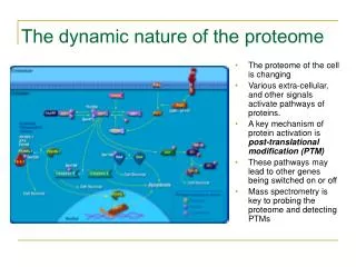
Olive oil (400 mL). +400 mL 25% TCA/Acetone. Precipitate redisolution in 4% SDS 25 mM DTT, 100 ºC. Precipitation MeOH/CH 3 Cl. SDS-PAGE-Silver staining. THE PROTEOME OF OLIVE SEEDS AND THE INVISIBLE PROTEOME OF OLIVE OILS AS DETECTED VIA COMBINATIONAL PEPTIDE LIGAND LIBRARY CAPTURE

E N D
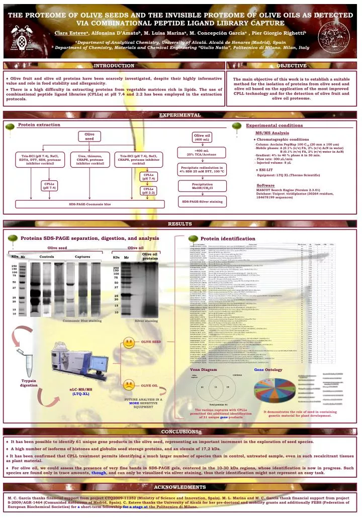
Olive oil (400 mL) +400 mL 25% TCA/Acetone Precipitate redisolution in 4% SDS 25 mM DTT, 100 ºC Precipitation MeOH/CH3Cl SDS-PAGE-Silver staining THE PROTEOME OF OLIVE SEEDS AND THE INVISIBLE PROTEOME OF OLIVE OILS AS DETECTED VIA COMBINATIONAL PEPTIDE LIGAND LIBRARY CAPTURE Clara Estevea, Alfonsina D’Amatob, M. Luisa Marinaa, M. Concepción Garcíaa , Pier Giorgio Righettib aDepartment of Analytical Chemistry, University of Alcalá. Alcalá de Henares (Madrid), Spain b Department of Chemistry, Materials and Chemical Engineering “Giulio Natta”, Politecnico di Milano. Milan, Italy INTRODUCTION OBJECTIVE The main objective of this work is to establish a suitable method for the isolation of proteins from olive seed and olive oil based on the application of the most improved CPLL technology and for the detection of olive fruit and olive oil proteome. ♠ Olive fruit and olive oil proteins have been scarcely investigated, despite their highly informative value and role in food stability and allergenicity. ♠ There is a high difficulty in extracting proteins from vegetable matrices rich in lipids. The use of combinational peptide ligand libraries (CPLLs) at pH 7.4 and 2.2 has been employed in the extraction protocols. EXPERIMENTAL Protein extraction Experimental conditions MS/MS Analysis Olive seed ♠ Chromatographic conditions • Column: Acclaim PepMap 100 C18 (20 mm x 100 µm) • Mobile phases: A (0.1% (v/v) FA, 2% (v/v) AcN in water) • B (0.1% (v/v) FA, 2% (v/v) water in AcN) • Gradient: 4% to 40 % phase A in 30 min. • - Flow rate: 300 µL/min • - Injected volume: 8 µL Urea, thiourea, CHAPS, protease inhibitor cocktail Tris-HCl (pH 7.4), NaCl, CHAPS, protease inhibitor cocktail Tris-HCl (pH 7.4), NaCl, EDTA, DTT, SDS, protease inhibitor cocktail ♠ ESI-LIT CPLLs (pH 7.4) • Equipment: LTQ XL (Thermo Scientific) CPLLs (pH 7.4) Software MASCOT Search Engine (Version 2.3.01) Database: Uniprot_viridiplantae (30264 residues, 184678199 sequences) CPLLs (pH 2.2) SDS-PAGE-Coomassie blue RESULTS Proteins SDS-PAGE separation, digestion, and analysis Protein identification Olive seed Olive oil Coomassie Blue staining Silver staining OLIVE SEED Venn Diagram GeneOntology Trypsin digestion OLIVE OIL nLC-MS/MS (LTQ-XL) FUTURE ANALYSIS IN A MORESENSITIVE EQUIPMENT The various captures with CPLLs permitted the additional identification of 31 unique geneproducts It demonstrates the role of seed in containing genetic material for plant development. CONCLUSIONS ♠ It has been possible to identify 61 unique gene products in the olive seed, representing an important increment in the exploration of seed species. ♠ A high number of isoforms of histones and globulin seed storage proteins, and an oleosin of 17,2 kDa. ♠ It has been confirmed that CPLL treatment permits identifying a much larger number of species than in control, untreated sample, even in such recalcitrant tissues as plant material. ♠ For olive oil, we could assess the presence of very fine bands in SDS-PAGE gels, centered in the 10-30 kDa regions, whose identification is now in progress. Such species are found only in trace amounts, though, and can only be visualized via silver staining, thus their identification might not represent an easy task. ACKNOWLEDMENTS M. C. García thanks financial support from project CTQ2009-11252 (Ministry of Science and Innovation, Spain). M. L. Marina and M. C. García thank financial support from project S-2009/AGR-1464 (ComunidadAutónoma of Madrid, Spain). C. Esteve thanks the University of Alcalá for her pre-doctoral and mobility grants and additionally FEBS (Federation of European Biochemical Societies) for ashort-term fellowshipfor a stage atthe Politecnico di Milano.