Download
1 / 39
510 likes | 1.64k Vues
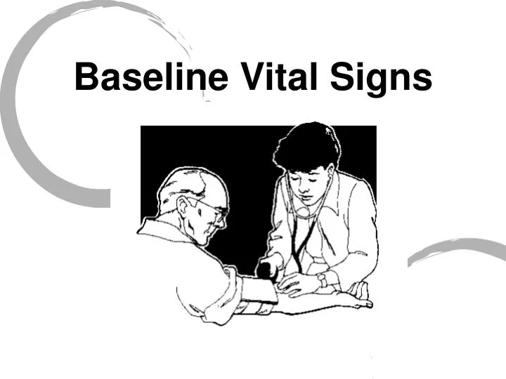
Baseline Vital Signs. Baseline Vital Signs. • Key signs used to evaluate a patient’s condition • First set is known as baseline vitals • Repeated vital signs compared to the baseline • Need at least 2 sets of vitals to show trending. Baseline Vital Signs. PRBELLS Pulse Respiration

E N D
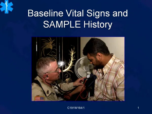
Baseline Vital Signs • Key signs used to evaluate a patient’s condition • First set is known as baseline vitals • Repeated vital signs compared to the baseline • Need at least 2 sets of vitals to show trending
Baseline Vital Signs • PRBELLS • Pulse • Respiration • Blood pressure • Eyes • Lung sounds • LOC - Level of Consciousness • Skins
Pulse • Rate – Number of beats in 30 seconds x 2 – Number of beats in 15 seconds x 4 • Rhythm – Regular or irregular • Quality – Bounding, strong, or weak (thready)
Normal Pulse Rates Adults 60 to 100 beats/min Children 70 to 150 beats/min Infants 100 to 160 beats/min Fast = Tachycardia - over 100 in adults Slow= Bradycardia - under 60 in adults
Respirations • Effort – Normal or Labored • Noisy respiration – Normal, stridor, wheezing, snoring, gurgling • Depth – Shallow or deep • Rate – Number of breaths in 30 seconds x 2 • Rhythm – Regular or irregular • Quality – Character of Breathing
Respiratory Rates Adults 12 to 20 breaths/min Children 15 to 30 breaths/min Infants 25 to 50 breaths/min
Common Terms • Bradypnea= slow breathing • Tachypnea= fast breathing • Eupnea= normal breathing • Apnea = no breathing
Blood Pressure • A drop in blood pressure may indicate: – Loss of blood – Loss of vascular tone – Cardiac pumping problem
Measuring Blood Pressure • Diastolic – Pressure during relaxing phase of the heart’s cycle • Systolic – Pressure during contraction • Measured as millimeters of mercury (mm Hg) • Recorded as systolic/diastolic
Auscultation of Blood Pressure 1. Place cuff on patient’s arm. 2. Palpate brachial artery and place stethoscope. 3. Inflate cuff until you no longer hear pulse sounds. 4. Continue pumping to increase pressure by an additional 20 mm Hg. 5. Note the systolic and diastolic pressures as 6. you let air escape slowly. 7. Korotkoff Sounds 8. 1st beat you hear is systolic 9. Last beat you hear is diastolic 10.As soon as pulse sounds stop, open the valve and release the air quickly.
Palpation of Blood Pressure • Secure cuff. • Locate radial pulse. • Inflate to about 200 mm Hg. • Release air until pulse is felt. • Method only obtains systolic pressure.
Normal Ranges of Blood Pressure Infants (newborn to 1 year) 50 to 95(systolic) Children (1 to 8 years) 80 to 110 mm Hg (systolic) Adults 90 to 140 mm Hg (systolic)
Pupil Assessment • P - Pupils • E - Equal • A - And • R - Round • R - Regular in size • L - React to Light
Abnormal Pupil Reactions • Fixed with no reaction to light • Dilate with light and constrict without light…that’s a brain problem! • React sluggishly • Unequal in size • Unequal with light or when light is removed
Level of Consciousness Name? Date? Place? Problem?
Lung Sounds Types of Lung Sounds • Lung sounds are typically broken down into three categories: • Normal (vesicular) • Decreased or absent • Abnormal (adventitious)
Lung Sounds • There are several types of abnormal lung sounds: • Wheezes are caused by air flowing rapidly through narrowed airway passages • Rhales are small bubbling or fine clicking sounds made when air is forced into collapsed alveoli and/or in the presence of fluid in the alveoli and/or bronchioles. • Rhonchi are low-pitched, sonorous, rumbling, bubbling or gurgling sounds. • Pleural rub, or friction rub, occurs when there is fluid in the pleural space between the lung tissue and the interior chest wall. There is commonly a grating, rubbing type of sound as the visceral (lung) and parietal (chest wall) pleura rub against each other.
Reassessment of Vital Signs • Reassess stable patients every 15 minutes.• Reassess unstable patients every 5 minutes
Patient History SAMPLE • S - Signs & symptoms • OPQRST • A - Allergies • M - Medications • P - Past medical history • L - Last oral intake • E - Events leading to incident
S - Signs & Symptoms OPQRST O - Onset When & How did the symptom begin? P - Provokes/Palliates What makes the symptom worse? What makes the symptom better?
S - Signs & Symptoms OPQRST Q - Quality • How would describe the pain?/What does the pain feel like? • DO NOT lead the patient R - Region/Radiation • Where is the pain? • Does the pain travel anywhere else?
S - Signs & Symptoms OPQRST S - Severity • How bad is the pain? Scale of 1-10 T- Time • How long have you had the symptom?
A - Allergies • Medications • Foods • Environment
M- Medications Are you taking any? When did you last take your medication? What are they? What are they for? May I see them? May we take them with us?
P - Previous Medical History • Pertinent • Related to this complaint • Complicating factor
L - Last Oral Intake Food and/or Drink? What? When?
E - Events leading up to the incident What happened? When?