Download
1 / 50
550 likes | 741 Vues
Anatomy of Arm. Dr. Fadel Naim Orthopedic Surgeon Islamic University Faculty of Medicine. Veins of the Arm. Two sets of veins, superficial and deep , anastomose freely with each other. The superficial veins are in the subcutaneous tissue The deep veins accompany the arteries .

E N D
Anatomy of Arm Dr. Fadel Naim Orthopedic Surgeon Islamic University Faculty of Medicine
Veins of the Arm • Two sets of veins, superficial and deep, anastomose freely with each other. • The superficial veins are in the subcutaneous tissue • The deep veins accompany the arteries. • Both sets of veins have valves • They are more numerous in the deep veins • The smooth muscles of the veins are innervated by sympathetic postganglionic nerve fibers
Superficial Veins • The two main superficial veins of the arm: • The cephalic vein • The basilic vein
Superficial Veins • The cephalic vein • Located in the subcutaneous tissue along the anterolateral surface of the proximal forearm and arm • Often visible through the skin • Passes superiorly between the deltoid and pectoralis major in the deltopectoral groove • It empties into the termination of the axillary vein in the deltopectoral triangle
Superficial Veins • The basilic vein • located in the subcutaneous tissue and passes on the medial side of the inferior part of the arm • Often it is also visible through the skin. • Near the junction of the middle and inferior thirds of the arm, the basilic vein penetrates the brachial (deep) fascia and runs superiorly into the axilla • Where it merges with the accompanying veins of the brachial artery to form the axillary vein
Deep Veins of the Arm • The brachial veins: • The paired veins accompanying the brachial artery • Their frequent connections encompass the artery, forming an anastomotic network within a common vascular sheath. • The pulsations of the brachial artery help move the blood through this venous network. • Begin at the elbow by union of the accompanying veins of the ulnar and radial arteries. • End by merging with the basilic vein to form the axillary vein.
Venipuncture of the Upper Limb • Because of the prominence and accessibility of the superficial veins of the upper limb, they are commonly used for venipuncture • These veins may be embedded with the subcutaneous tissue (fat), making them difficult to see • By applying a tourniquet to the arm, the venous return is occluded and the veins distend and are usually visible and/or palpable.
Venipuncture of the Upper Limb • The median cubital vein is commonly used for venipuncture for: • Drawing blood • Inserting a catheter for right cardiac catheterization • The dorsal venous network and the cephalic and basilic veins arising from it are commonly used for intravenous feeding
Lymphatic Drainage of the Upper Limb • Superficial lymphatic vessels • Arise from lymphatic plexuses in: • The digits • The palm • The dorsum of the hand • Ascend mostly with superficial veins • Deep lymphatic vessels: • Less numerous than superficial vessels • Accompany the major deep veins in the upper limb • Terminate in the humeral group of axillary nodes.
Lymphatic Drainage of the Upper Limb • Some vessels accompanying the basilic vein enter the cubital nodes, located: • proximal to the medial epicondyle • medial to the basilic vein. • Efferent vessels from these lymph nodes ascend in the arm and terminate in the humeral (lateral) axillary lymph nodes. • Most lymphatic vessels accompanying the cephalic vein cross the proximal part of the arm and anterior aspect of the shoulder to enter the apical group of axillary nodes; however, some vessels previously enter the deltopectoral nodes.
Lymphangitis, Lymphadenitis, and Lymphedema • Lymphangitis: • The inflammation of lymphatic vessels • Lymphadenitis: • The inflammation of lymph nodes • These conditions may occur when the lymphatic system is involved in the spread (metastasis) of cancer cells or infection
Fascial compartments of the Upper Limb • The brachial fascia: • A sheath of deep fascia encloses the arm like a sleeve • It is continuous superiorly with the pectoral and axillary layers of fascia. • It is attached inferiorly to the epicondyles of the humerus and the olecranon of the ulna and is continuous with the antebrachial fascia • Intermuscular septa: • The medial and lateral intermuscular septa extend from the deep surface of the brachial fascia to the medial and lateral supracondylar ridges of the humerus • Dividing the arm into • Anterior (flexor) fascial compartment • Posterior (extensor) fascial compartment • Each of which contains muscles serving similar functions, nerves, and blood vessels that supply
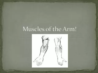
Lateral • head • Long • head • Triceps • brachii Posterior compartment of arm (extends elbow); innervation: radial nerve • Medial • head • Humerus • Extensors • Flexors • Brachialis • Others • Short head • Biceps brachii • Long head • (a) • (a) Muscles of the arm Anterior compartment of arm (flexes elbow); innervation: musculocutaneous nerve
Muscles of the Arm • 4 arm (brachial) muscles: • 3 flexors in the anterior compartment • Supplied by the musculocutaneous nerve • Biceps brachii • Brachialis • Coracobrachialis • 1 extensor in the posterior compartment • Triceps brachii • Supplied by the radial nerve.
BICEPS BRACHII • ORIGIN • Long head: • supraglenoid tubercle of scapula. • Short head: • coracoid process of scapula with coracobrachialis • INSERTION • posterior border of bicipital tuberosity of radius (over bursa) • bicipital aponeurosis to deep fascia and subcutaneous ulna • ACTION • Supinator of the forearm • Flexion of the elbow • weakly flexes shoulder • NERVE • Musculocutaneous nerve (C5, 6) (from lateral cord)
The tendon of the long head of the biceps crosses the head of the humerus within the cavity of the glenohumeral joint • The tendon, surrounded by synovial membrane, descends in the intertubercular groove of the humerus
The bicipitoradial bursa separates the biceps tendon from--and reduces abrasion against--the anterior part of the radial tuberosity.
When the elbow is extended • The biceps is a simple flexor of the forearm • When the elbow is flexed and more power is needed against resistance • The biceps is the primary (most powerful) supinator of the forearm • When right-handed persons drive a screw into hard wood • Inserting a corkscrew and pulling the cork from bottle. • The biceps barely operatesduring flexion of the prone forearm. • To test the biceps: • The ebow joint is flexed against resistance when the forearm is supinated. • If acting normally, the muscle forms a prominent bulge on the anterior aspect of the arm that is easily palpated.
Biceps Tendinitis • The tendon of the long head of the biceps, enclosed by a synovial sheath, moves back and forth in the intertubercular groove of the humerus. • Wear and tear of this mechanism is a common cause of shoulder pain. • Inflammation of the tendon (biceps tendinitis), usually the result of repetitive microtrauma, is common in sports involving throwing and use of a racquet • A tight, narrow, and/or rough intertubercular groove may irritate and inflame the tendon, producing tenderness and crepitus
Rupture of the Tendon of the Long Head of the Biceps • Often as the result of prolonged tendinitis that weakens it. • May result from repetitive overhead motions, such as occurs in swimmers • May result from forceful flexion of the arm against excessive resistance, as occurs in weight lifters • Usually the tendon is torn from its attachment to the supraglenoid tubercle of the scapula. • Commonly dramatic and is associated with a snap or pop. • The detached muscle belly forms a ball near the center of the distal part of the anterior aspect of the arm ("popeye deformity")
BRACHIALIS • ORIGIN • Anterior lower half of humerus • medial and lateral intermuscular septa • INSERTION • Coronoid process and tuberosity of ulna • ACTION • Flexes elbow • NERVE • Musculocutaneous nerve (C5, 6) ( from lateral cord). • Also small supply from radial nerve (C7)
The brachialis is the main flexor of the forearm • Flexes the forearm in all positions and during slow and quick movements. • When the forearm is extended slowly, the brachialis steadies the movement by slowly relaxing (picking up and put down a teacup carefully) • The brachialis always contracts during flexion of the elbow joint and is primarily responsible for maintaining flexion • Because of its many functions, it is regarded as the workhorse of the elbow flexors
CORACOBRACHIALIS • ORIGIN • Coracoid process of scapula with biceps brachii • INSERTION • Upper half medial border of humerus • ACTION • Flexes and weakly adducts arm • NERVE • Musculocutaneous nerve (C5, 6, 7) (from lateral cord)
Long head Biceps brachii Coracobrachialis Short head Biceps tendon Aponeurosis of biceps brachii
Acromion process Coracoid process Humerus Coracobrachialis Musculocutaneous n. Brachialis Radius Ulna
TRICEPS • ORIGIN • Long head: • infraglenoid tubercle of scapula. • lateral head: • upper half posterior humerus (linear origin). • medial head: • lies deep on lower half posterior humerus inferomedial to spiral groove and both intermuscular septa • INSERTION • Posterior part of upper surface of olecranon process of ulna and posterior capsule • ACTION • Extends elbow • Long head stabilizes shoulder joint • medial head retracts capsule of elbow joint on extension • NERVE • Radial nerve (C7, 8) (from posterior cord ), four branches
The triceps is the main extensor of the elbow joint. • Aids in extension and adduction of the arm. • Long head of the triceps helps stabilize the adducted glenohumeraljoint by serving as a shunt muscle, resisting inferior displacement of the head of the humerus. • Just proximal to its distal attachment is a friction-reducing subtendinous olecranon bursa between the triceps tendon and the olecranon. • To test the triceps lesion • the arm is abducted 90 ° and then the flexed forearm is extended against resistance • If acting normally, the triceps can be seen and palpated.
Deltoid (cut) Long head Triceps brachii Lateral head Olecranon Anconeus
Suprascapular nerve Dorsal scapular nerve Axillary nerve Lateral head Long head Triceps brachii Radial nerve Medial head Anconeus
Humeral Shaft Fracture • Fracture above the level of pectoralis major • proximal fragmentabduct and rotate internally due to the action of the rotator cuff • Fracture above the deltoid and below pectoralis major • deltoid pulls the distal fragment laterally, pectoralis major pulls the proximal fragment medially • Fracture below deltoid • proximal fragment abducts due to deltoid,distal fragment pulled medially and proximally by biceps/ brachialis and coracobrachialis
Arteries of the arm • Brachial • Deep (profunda) brachial • Anterior branch • Posterior branch • Superior ulnar collateral • Inferior ulnar collateral • Recurrent branches from: • The radial artery • Ulnar artery • Interosseous artery • These arteries anastomose with descending articular branches of the deep artery of the arm and the ulnar collateral arteries
Brachial Artery • The brachial artery provides the main arterial supply to the arm • The brachial artery, the continuation of the axillary artery • Begins at the inferior border of the teres major and ends in the cubitalfossa opposite the neck of the radius • Under cover of the bicipitalaponeurosis, the brachial artery divides into the radial and ulnar arteries
The brachial artery, superficial and palpable throughout its course • Lies anterior to the triceps and brachialis. • At first it lies medial to the humerus, and then anterior to it. • As it passes inferolaterally, the brachial artery accompanies the median nerve, which crosses anterior to the artery • During its course through the arm, the brachial artery gives rise to: • Many unnamed muscular branches • Humeral nutrient arteries, which arise from its lateral aspect
Deep Artery of the Arm • The largest branch of the brachial artery • Most superior origin • Accompanies the radial nerve through the radial groove and passes around the body of the humerus • Divides into anterior and posterior descending branches that participate in the arterial anastomoses around the elbow.
Nutrient Humeral Artery • Arises from the brachial artery around the middle of the arm and enters the nutrient canal on the anteromedial surface of the humerus. • The artery runs distally in the canal toward the elbow
Superior Ulnar Collateral Artery • This artery arises from the medial aspect of the brachial artery near the middle of the arm and accompanies the ulnar nerve posterior to the medial epicondyle of the humerus • It anastomoses with the : • posterior ulnar recurrent artery • inferior ulnar collateral artery
Inferior Ulnar Collateral Artery • Arises from the brachial artery approximately 5 cm proximal to the elbow crease • Passes inferomedially anterior to the medial epicondyle of the humerus • Joins the anastomoses of the elbow region by anastomosing with the anterior ulnar recurrent artery
Measuring Blood Pressure • Arterial blood pressure measurement using sphygmomanometer. • A cuff is placed around the arm and inflated with air until it compresses the brachial artery against the humerus and occludes it. • A stethoscope is placed over the artery in the cubital fossa, the pressure in the cuff is gradually released • The examiner detects the sound of blood beginning to spurt through the artery. • The first audible spurt indicates systolic blood pressure. • As the pressure is completely released, the point at which the pulse can no longer be heard is the diastolic blaod pressure.
Compressing the Brachial Artery • The best place to compress the brachial artery to control hemorrhage is near the middle of the arm • The brachial artery may be clamped distal to the deep artery of the arm without producing tissue damage • The anatomical basis for this is that the ulnar and radial arteries still receive sufficient blood through the anastomoses around the elbow • Ischemia of the elbow and forearm results from clamping of the brachial artery proximal to the deep artery of the arm for an extended period
Nerves of the Arm • 4 main nerves pass through the arm: • Median • Ulnar • Musculocutaneous • Radial • The median and ulnar nerves supply no branches to the arm.
Musculocutaneous Nerve • One of the terminal branches of the lateral cord • Supplies all the muscles in the anterior (flexor) compartment of the arm. • Begins opposite the inferior border of the pectoralis minor, pierces the coracobrachialis, and continues distally between the biceps and brachialis • After supplying all three of these muscles, it becomes the lateral cutaneous nerve of the forearm
Radial Nerve • The direct continuation of the posterior cord • Supplies all the muscles in the posterior compartment of the arm. • It enters the arm • Posterior to the brachial artery • Medial to the humerus • Anterior to the long head of the triceps • The radial nerve descends inferolaterally with the deep brachial artery and passes around the humeral body in the radial groove • Before entering the groove, it gives branches to the long and lateral heads of the triceps.
Radial Nerve • At the lateral border of the humerus, it pierces the lateral intermuscular septum and continues inferiorly in the anterior compartment of the arm between the brachialis and brachioradialis to the level of the lateral epicondyle of the humerus. • It then divides into deep and superficial branches • The deep branch is entirely muscular and articular in its distribution. • The superficial branch is entirely cutaneousin its distribution, supplying sensation to the dorsum of the hand and digits.
Median Nerve • Formed in the axilla by the union of a lateral root from the lateral cord and a medial root from the medial cord • It runs distally in the arm, initially on the lateral side of the brachial artery until it reaches the middle of the arm ,where it crosses to the medial side and contacts the brachialis. • The median nerve then descends to the cubital fossa, where it lies deep to the bicipital aponeurosis and medial to cubital vein. • The median nerve has no branches in the axilla or the arm, but it supplies articular branches to the elbow joint.
Ulnar Nerve • This is the larger of the two terminal branches of the medial cord • It passes distally, anterior to the triceps, on the medial side of the brachial artery • Around the middle of the arm it pierces the medial intermuscular septum with the superior ulnar collateral artery and descends between the septum and the medial head of the triceps. • The ulnar nerve passes posterior to the medial epicondyle and medial to the olecranon to enter the forearm • Posterior to the medial epicondyle where the ulnar nerve is referred to in lay terms as the “funny bone“ • The ulnar nerve has no branches in the arm, but it supplies articular branches to the elbow joint.