Download
1 / 40
400 likes | 497 Vues
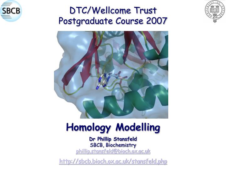
DTC/Wellcome Trust Postgraduate Course 2007. Dr Phillip Stansfeld SBCB, Biochemistry phillip.stansfeld@bioch.ox.ac.uk http://sbcb.bioch.ox.ac.uk/stansfeld.php. Homology Modelling. Contents. Introduce the process of homology modelling.

E N D
DTC/Wellcome Trust Postgraduate Course 2007 Dr Phillip Stansfeld SBCB, Biochemistry phillip.stansfeld@bioch.ox.ac.uk http://sbcb.bioch.ox.ac.uk/stansfeld.php Homology Modelling
Contents • Introduce the process of homology modelling. • Summarise the methods for predicting the structure from sequence. • Describe the individual steps involved in creating and optimising a protein homology model. • Outline the methods available to evaluate the quality of homology models. • Case Study – Modelling the Drug binding site of hERG.
Why Homology Model? • Solving protein structures is not trivial. • There are currently ~1.8 million known protein coding sequences. • But only ~44,000 protein structures in the PDB. • Even so, many of these structures are duplicates. • For Membrane Proteins structural data is even more sparse: • There are currently 304 membrane proteinstructures, of which only 142 are unique. www.rscb.org
Amino Acid Residues • Proteins are made up of amino acids, which are interconnected by peptide bonds. • There are 20 naturally occurring amino acids. • Amino acids may be subdivided by their individual properties.
From Sequence to Structure Primary Structure – Amino Acid Sequence DSSRRQYQEKYKQVEQYMSFHKLPADFRQKIHDYYEHRYQGKMFDEDSILGELNGPLREEIVNFNCR KLVASMPLFANADPNFVTAMLTKLKFEVFQPGDYIIREGTIGKKMYFIQHGVVSVLTGNKEMKLSDG SYFGEICLLTRGRRTASVRADTYCRLYSLSVDNFNEVLEEYPMMRRAFETYVAIDRLDRIGKKNSIL Secondary Structure Tertiary Structure Quaternary Structure What information can we get from a Sequence of amino acids?
Secondary Structure Prediction • The Secondary Structure of Proteins is Defined by the DSSP algorythm. • Amino acids classified as either α-helix (H), β-strand (S) or loop (C). • It is possible to extract structural information from amino acid sequence. • These prediction methods were initially proposed by Chou & Fasman in 1978. • They used a statistical method based on 15 known crystal structures. • Recent developments and an increase in structural information has improved these methods and they are currently ~80% accurate. PSI-Pred: http://bioinf.cs.ucl.ac.uk/psipred/psiform.html JPred: http://www.compbio.dundee.ac.uk/~www-jpred/
Transmembrane Helix Prediction • The amino acids at the centre of transmembrane helices are generally hydrophobic in nature. • Analysis of Hydropathicity can be used to predict the number of membrane spanning helices. • The analysis for the G-protein coupled receptor to the right suggests it has 7 TM helices. • The example used the Kyte & Doolittle scale. Hydropathy Plot http://expasy.org/tools/protscale.html
BLAST • How to find an appropriate template Structure for homology modelling… • Basic Local Alignment Search Tool • Used to search protein databases: • e.g. Non-redundant (nr) & SwissProt to find similar sequences. • Protein Data Bank (PDB) to find structures with similar sequences. • PSI- & PHI-blast are more advanced Blast methods. http://www.ncbi.nlm.nih.gov/blast/Blast.cgi
low high The Importance of Resolution • In X-ray crystallography it is not always possible to flawlessly resolve the crystal density of the protein of interest. • This results in a lower resolution structure. • The lower the resolution the more likely the structure is wrong. • The resolution of the template structure also reflects in the quality of the homology model. 4 Å 3 Å 2 Å 1 Å
Sequence Alignment • Aligns the sequence(s) of interest to that of the template structure(s). • Emboss may be used for two sequence, to generate a pairwise alignment & a percentage identity – ideally an identity of >50%: http://www.ebi.ac.uk/emboss/align/ • T-Coffee, Clustal & MUSCLE are popular methods for multiple sequence alignment. All may be found at : http://www.ebi.ac.uk/ • ESPRIPT is useful for formatting to creating black & white figures: http://espript.ibcp.fr/
Automated Homology Modelling If you are lazy there are servers that do the modelling for you! • Swiss Model http://swissmodel.expasy.org//SWISS-MODEL.html • Robetta http://robetta.bakerlab.org/ • 3D Jigsaw http://www.bmm.icnet.uk/servers/3djigsaw/ • Phyre http://www.sbg.bio.ic.ac.uk/phyre/ • EsyPred3D http://www.fundp.ac.be/sciences/biologie/urbm/bioinfo/esypred/ • CPHmodels http://www.cbs.dtu.dk/services/CPHmodels/
Modeller from modeller import * from modeller.automodel import * log.verbose() env = environ() env.io.atom_files_directory = './' a = automodel( env, alnfile = 'herg.ali', knowns = '1q5o', sequence = 'herg' ) a.starting_model= 1 a.ending_model = 1 a.make() >P1;1q5o structureX: 1q5o : 443 : A : 644 : A :::: DSSRRQYQEKYKQVEQYMSFHKLPADFRQKIHDYYEHRYQ-GKMFDEDSILGELNGPLRE EIVNFNCRKLVASMPLFANADPNFVTAMLTKLKFEVFQPGDYIIREGTIGKKMYFIQHGV VSVLTKGNKEMKLSDGSYFGEICLL--TRGRRTASVRADTYCRLYSLSVDNFNEVLEEYP MMRRAFETVAIDRLDRIGKKNSIL.* >P1;herg sequence: herg : 1 ::::::: YSGTARYHTQMLRVREFIRFHQIPNPLRQRLEEYFQHAWSYTNGIDMNAVLKGFPECLQA DICLHLNRSLLQHCKPFRGATKGCLRALAMKFKTTHAPPGDTLVHAGDLLTALYFISRGS IEILRGDVVVAILGKNDIFGEPLNLYARPGKSNGDVRALTYCDLHKIHRDDLLEVLDMYP EFSDHFWSSLEITFNLRDTN-MIP.* • Well regarded program for Homology/Comparative Modelling. • Current Version 9v2. http://www.salilab.org/modeller/ • Requires an Input file, Sequence alignment & Template structure. Sequence Alignment (*.ali) ATOM 1 N ASP A 443 -15.943 41.425 44.702 1.00 44.68 ATOM 2 CA ASP A 443 -15.424 42.618 45.447 1.00 43.15 ATOM 3 C ASP A 443 -14.310 43.306 44.686 1.00 41.81 ATOM 4 O ASP A 443 -14.298 44.528 44.539 1.00 42.61 etc... Template Structure (*.pdb) Input File (*.py)
How Does it Work? Glutamine Valine Change in Rotamer Amino acid Substitution Energy Minimisation Template Structure Initial Model (*.ini) Output Model(s) (*.B999*)
Modeller : Output • .log : log output from the run. • .B* : model generated in the PDB format. • .D* : progress of optimisation. • .V* : violation profile. • .ini : initial model that is generated. • .rsr : restraints in user format. • .sch : schedule file for the optimisation process.
Modeller Features & Restraints • Secondary Structure. Regions of the protein may be forced to be α-helical or β-strand. • Distance restraints. The distance between atoms may be restrained. • Symmetry. Proteinmultimers can be restrained so that all monomers are identical. • Disulphide Bridges. Two cysteine residues in the model can be forced to make a cystine bond. • Ligands. Ions, waters and small molecules may be included from the template. • Loop Refinement. Regions without secondary structure often require further refinement.
Structural Convergence • The catalytic triad of Serine, Aspartate and Histidine is found in certain protease enzymes. (a) Subtilisin (b) Chymotrypsin. • However, the overall structure of the enzyme is often different. • This is also important when considering ligand binding sites.
Modelling Ligand Interactions ATP Binding Site • Small molecules, waters and ions can be retained from the template structure. • It is possible to search for homologues based on the ligands they bind. • Experimental data, especially mutagenesis is very useful when modelling ligand binding sites. • Although the key residues may often remain, the overall structure of the protein may vary radically. • The presence of the ligand is also likely to alter the conformation of the protein. 1ATN 1E4G
Conformational States • The backbone structure of the model will be almost identical to that of the template. • Therefore the conformational state of the template will be retained in the resultant homology model. • This is important when considering the open or closed conformation of a channel… • … or the Apo versus bound state of a ligand binding site. Closed Open
Loop Modelling Issues with Loop Modelling • As loops are less restrained by hydrogen bonding networks they often have increased flexibility and therefore are less well defined. • In addition the increased mobility make looped regions more difficult to structurally resolve. • Proteins are often poorly conserved in loop regions. • There are usually residue insertions or deletions within loops. • Proline and Glycine resides are often found in loops – we’ll come back to this when discussing Model evaluation protocols.
Loop Modelling • There are two main methods for modelling loops: • Knowledge based: A PDB search for fragments that match the sequence to be modelled. • Ab initio: A first principles approach to predict the fold of the loop, followed by minimisation steps. • Many of the newer loop prediction methods use a combination of the two methods. • These approaches are being developed into methods for computationally predicting the tertiary structure of proteins. eg Rosetta. • But this is computationally expensive. • Modeller creates an energy function to evaluate the loop’s quality. • The function is then minimised by Monte Carlo (sampling), Conjugate Gradients (CG) or molecular dynamics (MD) techniques.
Predicting Sidechain Conformations • Networks of side chain contacts are important for retaining protein structure. • Sidechains may adopt a variety of different conformations, but this is dependent on the residue type. • For example a threonine generally adopts 3 conformations, whilst a lysine may adopt up to 81. • This is dependent backbone conformation of the residue. • The different residue conformations are known as rotamers. • Where a residue is conserved it is best to keep the side chain rotamer from the template than predict a new one. • Rotamer prediction accuracy is high for buried residues, but much lower for surface residues: • Side chains at the surface are more flexible. • Hydrophobic packing in the core is easier to handle than the electrostatic interactions with water molecules. (cytoplasmic proteins) • Most successful method is SCWRL by Dunbrack et al.: http://dunbrack.fccc.edu/SCWRL3.php
Model Evaluation Initial Options • For every model, Modeller creates an objective function energy term, which is reported in the second line of the model PDB file (.B*). • This is not an absolute measure but can be used to rank models calculated from the same alignment. The lower the value the better. • A Cα-RMSD (Root Mean Standard Deviation) between the template structure and models can also be used to compare the final model to its template. • A good Cα-RMSD will be less than 2Å.
Model Evaluation More Advanced Options • Procheck, PROVE, WhatIf: Stereochemical checks on bond lengths, angles and atomic contacts. • Ramachandran Plot is a major component of the evaluation. • Ensures that the backbone conformation of the model is normal. • Modeller is good on the whole, but sometimes struggles with residues found in loops. • RAMPAGE: β-strand left-handed helix Psi Dihedral Angle α-helix http://mordred.bioc.cam.ac.uk/~rapper/rampage.php Phi Dihedral Angle
Ramachandran Plot • The results of the ramachandran plot will be very similar to that of the template. • A Good template is therefore key! • Most residues are mainly found on the left-hand side of the plot. • Glycine is found more randomly within plot (orange), due to its small sidechain (H) preventing clashes with its backbone. • Proline can only adopt a Phi angle of ~-60° (green) due to its sidechain. • This also restricts the conformational space of the pre-proline residue. Peptide dihedral angles N
PROCHECK +----------<<< P R O C H E C K S U M M A R Y >>>----------+ | | | mgirk .pdb 2.5 104 residues | | | *| Ramachandran plot: 91.7% core 7.6% allow 0.3% gener 0.4% disall | | | *| All Ramachandrans: 15 labelled residues Backbone | *| Chi1-chi2 plots: 6 labelled residues Sidechain | | Main-chain params: 6 better 0 inside 0 worse | | Side-chain params: 5 better 0 inside 0 worse | | | *| Residue properties: Max.deviation: 16.1 Bad contacts: 10 | *| Bond len/angle: 8.0 Morris et al class: 1 1 3 | | | | G-factors Dihedrals: 0.10 Covalent: 0.29 Overall: 0.16 | | | | M/c bond lengths: 99.1% within limits 0.9% highlighted | *| M/c bond angles: 98.1% within limits 1.9% highlighted | | Planar groups: 100.0% within limits 0.0% highlighted | | | +----------------------------------------------------------------------------+ + May be worth investigating further. * Worth investigating further. Biotech Validation Suite: http://biotech.embl-ebi.ac.uk:8400/ Procheck: www.biochem.ucl.ac.uk/~roman/procheck/procheck.html
CASP • Critical Assessment of Structure Prediction. • A Biennial competition that has run since 1994. • The next competition will be in 2008 (CASP8) • http://predictioncenter.org/ • Its goal is to advance the methods for predicting protein structure from sequence. • Protein structures yet to be published are used as blind targets for the prediction methods, with only sequence information released. • Competitors may use Homology Modelling, Fold recognition or Ab Initio structural prediction methods to propose the structure of the protein.
Pymol • A powerful visualisation and picture generation tool for protein and DNA. • Two windows • Graphical User Interface (GUI) • Pymol Viewer • Both Text and Mouse driven. • Website: http://pymol.sourceforge.net/ • More Info & Tutorials: http://www.pymolwiki.org/ A-Action S-Show H-Hide L-Label C-Colour Sequence Viewer
Pymol Primary Uses • Visualisation of Macromolecular Structures. • High quality image generation capabilities (~1/4 of published images). • Structural alignment of two structures in three dimensional space. • Single amino acid mutagenesis. • Investigating Protein-Ligand interactions. • Assessing multiple-frame simulation data – not as robust as VMD.
Homology ModellingCase Study:Drug Binding Site of the hERG Potassium Channel
hERG Subunit Topology Selectivity Filter Turret Helix Voltage Sensor Domain Extracellular + + + S4 + + + _ _ S2 _ S1 _ S3b _ Pore Helix S5 S6 Pore Domain _ S3a Intracellular N-Terminal Domain C-Terminal Domain
Templates for Homology Modelling Filter KcsA KirBac1.1 MthK KvAP
Amino Acids involved in Drug Binding Selectivity Filter P V625 S624 G648 T623 Y652 F656 S5 V659 S6 Drug Access
Closed and Open State hERG KcsA Based KvAP Based
Ligand Docking to hERG KcsA Based - Closed KvAP Based - Open
Combining Individual Template Structures into a Complete Model 1ORS 1ORQ 1EYW 1Q5O
Predicting Conformational Changes Side Below Morph Server: http://www.molmovdb.org/cgi-bin/submit.cgi
Summary • Homology Modelling is a valuable tool for structural biologists. • There are five main stages: • Identify an appropriate template structure(s). • Create a Sequence alignment. • Perform the homology modelling. • Analyse and Evaluate the quality of the model. • Refinement. • It is important to take time when constructing a model – Crystallography is difficult & time consuming! • A model should not be rushed and should be fully checked! http://weblearn.ox.ac.uk/site/medsci/bioch/postgrad/compbio/2007dec/ps/
Practical Session • The notes and files for the Practical session can be found at: http://weblearn.ox.ac.uk/site/medsci/bioch/postgrad/compbio/2007dec/ps/ Or http://sbcb.bioch.ox.ac.uk/stansfeld.php/ • The file name is dtc_homology.tar • Untar the file in your home directory using: • tar cvf dtc_homology.tar • This will produce a folder called DTC, which contains three Exercises. • There are also two word documents: • Homology_Modelling_Practical_07.doc – Details of the practical. • Homology_Practical_Notes.doc – For your results. • If you need any help please let me know.
Practical Session • Details of the Three Exercises: • (a) Online Sequence Alignment Generation. (b) Homology Modelling a Monomer. (c) Evaluation & Visualisation. (d) Refinement. • (a) Retrieve the Sequence of interest. (b) Find a Suitable Template. (c) Modeller Sequence Alignment Generation. (d) Homology Modelling a Dimer. • (a) Homology Modelling a Tetramer with Ligands. (b) Structural Alignment of Template to Model. (c) Visualising Ligand Binding Sites. (d) Computational Mutagenesis.