Download
1 / 27
320 likes | 465 Vues
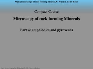
Compact Course Microscopy of rock-forming Minerals Part 4: Biotite, Mus c ovite, Chlorite, Turmaline, Titanite, Epidote, Stilpnomelane. Biotite Formula : K(Mg,Fe) 3 [(OH) 2 | AlSi 3 O 10 ] Symmetry : monoclinic n : 1,522 – 1,697 n : 0,04 – 0,08 2V : small, pseudo-uniaxial

E N D
Compact Course Microscopy of rock-forming Minerals Part 4: Biotite, Muscovite, Chlorite, Turmaline, Titanite, Epidote, Stilpnomelane
Biotite Formula : K(Mg,Fe)3[(OH)2|AlSi3O10] Symmetry : monoclinic n : 1,522 – 1,697 n : 0,04 – 0,08 2V : small, pseudo-uniaxial max. I. F. (30μm) : high, II. – IV. order Observations: Abundant sheet silicate (in volcanic, plutonic, metamorphic rocks). Strongly pleochroic from brown to green, with red hues. Mg-rich phlogopite almost colourless, only distinguished from white micas by 2V angle. Typical (for ALL micas !!) birds eyes structures. Pseudo-uniaxial through stacked twins ( c). In plutonic and metamorphic rocks typically with pleochroic halos around e.g. zircon inclusions Phlogopite
Weak Absorption strong Absorption Direction of polarizor strong Absorption 0,5 mm Biotite • Observations: • vertical to c:pseudo-uniaxial • parallel c • Note: no pleochroism, no traces of cleavage planes, • no birds eyes .......in plane vertical c ! Note! Pleochroism of biotite provides a good test for the polarization direction of thepolarizers at your microscope !
0,2 mm Biotite Observations: Bird's Eyes are typical for ALL micas, they are an artefact of thin section preparation.
0,5 mm Biotite, Replacement by Chlorite Observations: Retrograde replacement of biotite by chlorite is typical in many rocks. Replacement often is observed to ingress the biotite grains along their cleavage. Complete replacement of biotite by chlorite is possible.
Vertical c Parallel c 0,2 mm Biotite,Replacemente by chlorite and rutile Observations: Replacing Ti-bearing biotie by chlorite leaves exces Ti that cannot be incorporated in chlorite. This results in the formation of rutile needles (sagenite twins, ~ 60°).
0,3 mm Biotite,Replacement with formation of titanite (sphene) Observations: Replacement of Ti-rich biotite resulted in the formation of titanite (pseudonyme: sphene, CaTi[O|SiO4]).
Muscovite Formula : KAl2[(OH)2|AlSi3O10] Symmetry : monoclinic n : 1,552 – 1,624 n : 0,036 – 0,054 2V : 35° - 50° max. I. F. (30μm) : II. – III. order Observations: Colourless, all other features similar to biotite. Bird's Eyes! Orientation vertical c difficult to regognise: no birds eyes, no traces of cleavage, only colourless, featureless grains.
1 mm Muscovite • Observations: • colourless, no pleochroism, otherwise similar to biotite. • // c • vertical c • Biaxial, negative
Chlorite Formula : complex sheet silicate Symmetry : monoclininc n : 1,56 – 1,68 n : 0 – 0,01 2V : 0° - 30° max. I. F. (30μm) : grey I Observations: Shape and cleavage typical for sheet silicates. Colour faint green, slighty pleochroic. Anomalous interference colours in crystals with low birefringence. Typical for low-grade metamorphic shists, often replacing biotite during retrogression
0,3 mm Chlorite Observations: Chlorites have faint green colours, faint pleochrosim, low interferecne colours, anomalous interference colours (blueish). Anomalous colours and optical character (l') allows a simple determination of chlorite compositions (Mg/Fe ratio).
0,2 mm Chlorite Oservations: Colourless in normal light Anomalous brownish interference colour. negative elongation Rhipidolite
Turmaline Formiula : complex group of boro-silicates Symmetry : trigonal n : 1,620 – 1,692 n : 0,019 – 0,035 2V : - max. I. F. (30μm) : II. Order Observations: Typical trigonal cross sections and zonations. Coloured varieties are strongly pleochroic, strongly absorbing directions are vertical to elongation (in contrast to biotite!)
ne weak absorption no strong absorption Direction of polarizor 0,5 mm Turmaline • Observation: • cross section with vertical c trigonal, uniaxial negative. • longitudinal cross section • Note! Cross section with vertical c does not show pleochroism !
Titanite (or „sphene“) Formula : CaTi[O|SiO4] Symmetrye : monoclinic n : 1,921 – 2,08 n : 0,11 – 0,16 2Vz : 23° - 40° max. I. F. (30μm) : very high, white highst order Observations: Easy to recognise by a combination of (1) very high birefringence, (2) very high interference colours (white) and high „chagrain“ = granular surface texture
1 mm Titanite, typical crystal shapes („Sphene“) Observations: Easy to recognise by a combination of (1) very high birefrngence, (2) very high interference colours (white) and high „chagrins“ = granular surface texture Small axial angle, very strong dispersion of axial angle r>>v. Note: at right margin of image : garnet for comparison of relief and chagrins!
0,2 mm Titanite, typical bladed shape („Sphene“) Observation: Idiomorphic crystals in fine-grained matrix, twins. Note: very high abundance of small inclusions in matrix make it appear with high chagrins. Therefore,titanite which also has high chagrins can be more easly observed under crossed polarizers.
0,3 mm Titanite Observations: Shapes similar to ant-eggs. Note pleochroic haloes in amphibole: Sphene is rich in Th and U ! Note, pleochroic haloes only visible in Mg-Fe minerals, never in quartz or feldspars or other felsic minerals.
1 mm Titanite Observations: Titanite with inclusions of idiomorphic plagioclase. Titanite grows at the expense of biotite, resulting in pale haloes with no biotite and plagioclase dominating.
Epidote Formula : Ca2(Al,Fe)Al2[O|OH|SiO4|Si2O7] Symmetrie : monoclinic n : 1,68 – 1,78 n : 0,005 – 0,048 2Vz : 65° - 115° max. I. F. (30μm) : III. Order Observations: Anormalous „Super“- interference colours, Zonation! Fe3+-rich compositions are green. Typical mineral of metamorphic rocks (greenshist facies), often formed retrograde. Rarely magmatic in intrusive rocks.
0,3 mm Epidote Observations: Epidote with high refractive index often show „excessive“ interference colours. These are more intense and more brilliant than normal interference colours of the same order.
[010] b (010) 0,5 mm Epidote Observations: Strongly zoned crystal, cut (010). Symmetry is monoclinic, therefore the position of the indicatirx rotates freely around [010] axes (b-axis) as a function of composition (Fe-content). Note also good cleavage parallel (001).
0,5 mm Epidot Observations: Zonation twins, „excessive“ interference colours (canary yellow). When cut near isotropic plane, anomalous blueish interference colours.
1 mm Epidote Observations: Epidote and blue amphibole in tight folding pattern.
Stilpnomelane Formula : complex chain silicate with sheet-like structure Symmetry : monoclinic n : 1,54 – 1,75 n : 0,03 – 0,11 2V : small, pseudo-uniaxial ax. I. F. (30μm) : II. –V. order Observations: Very similar in most properties to biotite, except Bird's Eyes. Typical for high-P, low-T metamorphic rocks (blue shists).
0,5 mm Stilpnomelane Observations: Similar to biotite but no Bird's Eyes!!
Melatope (Achsenausstichpunkte) für blaues und rotesLicht Melatope und Isogyren für blaues Licht (isotrope Raumrichtungen und Auslöschungslagen für Blau), die restlichen Wellenlängen mischen sich zu Rot Melatope und Isogyren für rotes Licht (isotrope Raumrichtungen und Auslöschungslagen für Rot), die restlichen Wellenlängen mischen sich zu Blau Warum ist die Farbverteilung in Diagonalstellung umgekehrt zur tatsächlichen Dispersion?