Download
1 / 38
440 likes | 1.27k Vues
Muscular dystrophy. Dr. Derakhshandeh. Muscular dystrophy. Muscular dystrophy (MD) is a group of rare inherited muscle diseases in which muscle fibers are unusually susceptible to damage. Muscles, primarily voluntary muscles, become progressively weaker

E N D
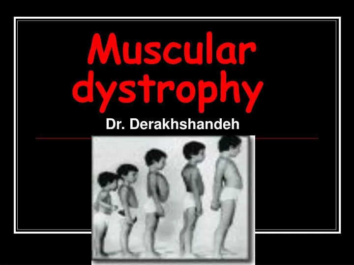
Muscular dystrophy Dr. Derakhshandeh
Muscular dystrophy • Muscular dystrophy (MD) is a group of rare inherited muscle diseases in which muscle fibers are unusually susceptible to damage. • Muscles, primarily voluntary muscles, become progressively weaker • In some types of muscular dystrophy, heart muscles, other involuntary muscles and other organs are affected.
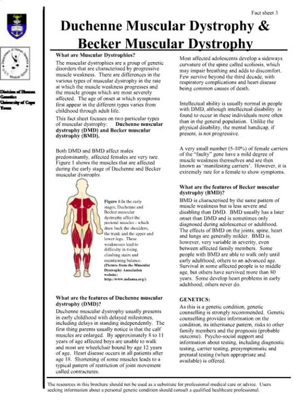
Duchenne's muscular dystrophy (Xp21.2) • The types of muscular dystrophy that are due to a genetic deficiency of the protein dystrophin are called dystrophinopathies. • Duchenne's muscular dystrophy is the most severe form of dystrophinopathy. • It occurs mostly in young boys and is the most common form of MD that affects children.
Dystrophin • a large (427 kD) cytoskeletal protein • localizes to the inner face of the skeletal muscle membrane • structure with an actin-binding domain at the amino terminus (N) • The carboxy-terminal domains associate with a large transmembrane complex of glycoproteins • directly bind with elements of the extracellular • Dystrophin: likely plays a critical role in establishing connections between the internal, actin-based cytoskeleton and the external basement membrane • Its absence may lead to increased membrane fragility
Duchenne's muscular dystrophy • Difficulty getting up from a lying or sitting position • Weakness in lower leg muscles, resulting in difficulty running and jumping • Waddling gait • Mild mental retardation, in some cases
In the late stages of muscular dystrophy, fat and connective tissue often replace muscle fibers.
Orthopaedic management of patients with Duchenne's muscular dystrophy
Duchenne's muscular dystrophy • X-linked inheritance Prevalence 0.003-0.05/1,000 total • Signs and symptoms of Duchenne's usually appear between the ages of 2 and 5 • It first affects the muscles of the pelvis, upper arms and upper legs. • By late childhood, most children with this form of muscular dystrophy are unable to walk.
Most die by their late teens or early 20s, often from pneumonia, respiratory muscle weakness or cardiac complications. • Some people with Duchenne's MD may exhibit curvature of their spine (scoliosis).
Becker's muscular dystrophy • This type of muscular dystrophy is a milder form of dystrophinopathy. • It generally affects older boys and young men, and progresses more slowly, usually over several decades. • Signs and symptoms of Becker's MD are similar to those of Duchenne's. • The onset of the signs and symptoms is generally later, from age 2 to 16.
L A B C D E F G H L ~95% of deletions can be detected in males using multiplex PCR
MAPH • Detection of deletions/duplication mutations in Duchenne Muscular Dystrophy using: Multiplex Amplifiable Probe Hybridisation (MAPH)
MAPH • Although ~95% of deletions can be detected in males using multiplex PCR • other methods must be used to determine duplications, as well as the carrier status of females • The most commonly applied methods are quantitative multiplex PCR and quantitative Southern blotting • The drawback of quantitative multiplex PCR is that often not all mutations are examined • meaning that small and rare mutations are missed
MAPH • Using high-quality Southern blots it is possible to perform a quantitative analysis and detect duplications • this technique is time consuming • it is difficult to exactly determine the duplication • it can be difficult to detect duplications in females and triplications will be missed Armour et al (Nucl.Acids Res. 2000)
system for analysing all 79 exons of the DMD gene for deletions and duplications • MAPH is based on a quantitative PCR of short DNA probes recovered after hybridisation to immobilized genomic DNA
1 ug of denatured genomic DNA is spotted on a small nylon filter • hybridized overnight in a solution containing one of the probe mixes • Following stringent washing the next day the filter is placed in a PCR tube • and a short PCR reaction is performed • This releases the specifically-bound probes into the solution • An aliquot of this is transferred to a second, quantitative PCR reaction
Myotonic dystrophy • This form of muscular dystrophy produces stiffness of muscles and an inability to relax muscles at will, as well as the muscle weakness of the other forms of muscular dystrophy. • The inability to relax muscles at will (myotonia) is found only in this type of muscular dystrophy.
Myotonic dystrophy • This form of MD can affect children, it often doesn't affect people until adulthood. • It can vary greatly in its severity. • Muscles may feel stiff after using them. • Progression of this form of MD is slow.
Myotonic dystrophy • Besides myotonia, signs and symptoms of adult-onset myotonic dystrophy may include: • Weakening of voluntary muscles • the muscles of the feet, hands, lower legs and forearms. • Weakening of head, neck and face muscles, which may result in the face having a hollow, drooped appearance. • Weakening of muscles involved in breathing and swallowing. • Weaker breathing muscles may result in less oxygen intake and fatigue. • Weaker swallowing muscles increase the risk of choking.
Myotonic dystrophy • Difficulty sleeping well at night and daytime sleepiness, and inability to concentrate. • Clouding of the lenses of the eyes (cataracts). • Mild diabetes.
Rarely, infants have this form of muscular dystrophy, in which case it's called congenital myotonic dystrophy. Signs in infants include: Severe muscle weakness Difficulty suckling and swallowing Difficulty breathing
The other major types of muscular dystrophy are rare. They include: • Limb-girdle muscular dystrophy • Facioscapulohumeral muscular dystrophy • Congenital muscular dystrophy • Oculopharyngeal muscular dystrophy • Distal muscular dystrophy • Emery-Dreifuss muscular dystrophy
Limb-girdle muscular dystrophy • Muscles usually affected first by this form of muscular dystrophy include: • Hips • Shoulders • This form then progresses to the arms and legs, though progression is slow. • Limb-girdle MD usually begins in the teen or early adult years.
Facioscapulohumeral muscular dystrophy • Also known as Landouzy-Dejerine disease, this form involves progressive muscle weakness, usually in this order: • Face • Shoulders • Abdomen • Feet • Upper arms • Pelvic area • Lower arms • When someone with facioscapulohumeral MD raises his or her arms, the shoulder blades may stick out like wings. • Progression of this form is slow, with some spurts of rapidly increasing weakness. • Onset usually occurs during the teen to early adult years.
Congenital muscular dystrophy • Signs of congenital MD may include: • General muscle weakness • Joint deformities • This form is apparent at birth and progresses slowly. • A more severe form of congenital MD called “Fukuyama” type congenital muscular dystrophy may involve severe mental and speech problems as well as seizures.
Oculopharyngeal muscular dystrophy • The first sign of this type of muscular dystrophy is usually drooping of the eyelids, followed by weakness of the muscles of the eye, face and throat, resulting in difficulty swallowing. • Progression is slow. • Signs and symptoms first appear in adulthood, usually in a person's 40s, 50s or 60s.
Distal muscular dystrophy • This group involves the muscles farthest away from the center of the body: • those of the hands, forearms, feet and lower legs. • The severity is generally less than for other forms of MD, and this form tends to progress slowly. • Distal MD generally begins in adulthood between the ages of 40 and 60.
Emery-Dreifuss muscular dystrophy • This rare form of muscular dystrophy usually begins in the muscles of the: • Shoulders • Upper arms • Shins • Emery-Dreifuss MD usually begins in the childhood to early teen years and progresses slowly.
Screening and diagnosis • A careful review of the family's history of muscle disease can help for a diagnosis. • Blood tests.: Damaged muscles release enzymes such as creatine kinase (CK) into the blood. High blood levels of CK suggest a muscle disease such as muscular dystrophy. • Electromyography.: A thin-needle electrode is inserted through the skin into the muscle to be tested. Electrical activity is measured as patient relax and as patient gently tighten the muscle. • Changes in the pattern of electrical activity can confirm a muscle disease. • The distribution of the disease can be determined by testing different muscles.
Screening and diagnosis • Ultrasonograph:High-frequency sound waves are used to produce precise images of tissues and structures within patients body. • An ultrasound is a noninvasive way of detecting certain muscle abnormalities, even in the early stages of the disease. • Muscle biopsy: A small piece of muscle is taken for laboratory analysis. • The analysis distinguishes muscular dystrophies from other muscle diseases. • Special tests can identify dystrophin and other markers associated with specific forms of muscular dystrophy.
Genetic testing • In the past, certain blood tests that are used to analyze DNA allowed some forms of muscular dystrophy to be diagnosed by identifying a particular mutation of the dystrophin gene. • Researchers are hoping that this test will soon become more widely available to the public.
Medications • Doctors prescribe medications to treat some forms of muscular dystrophy: • For myotonic dystrophy. The medications phenytoin), quinine may be used to treat the delayed muscle relaxation that occurs in myotonic dystrophy. • For Duchenne's muscular dystrophy. The anti-inflammatory corticosteroid medication “prednisone” may help improve muscle strength and delay the progression of Duchenne's MD.