Download
1 / 124
1.27k likes | 2.07k Vues
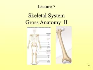
Anatomy of Skeletal Elements. The Musculoskeletal system. 206 bones grouped into the axial and appendicular skeletons 650 muscles approximately 40% of your body weight also divided into an axial and an appendicular division. Classification of Bones.

E N D
The Musculoskeletal system • 206 bones • grouped into the axial and appendicular skeletons • 650 muscles • approximately 40% of your body weight • also divided into an axial and an appendicular division
Classification of Bones • 6 types - based on anatomical classification • Long bones = greater length than width • Short bones = cube-shaped, spongy bone except at surface • Flat bones = two parallel plates of compact bone sandwiching spongy bone layer
Irregular bones = cannot be grouped • Sesamoid bones = develop in tendons where there is considerable friction, tension and stress • Sutural bones = located within joints between cranial bones
Bone Markings (surface features) • Used to identify specific elevations, depressions, and openings of bones • Bone markings provide distinct and characteristic landmarks for orientation and identification of bones and associated structures.
Bony Processes • Depressions and openings • Fissure – narrow slit • Foramen – hole for nerves, blood vessels • Fossa – cuplike depression • Sulcus – furrow on a bone surface, contains a nerve or blood vessel • Meatus – tubelike opening • Processes – projection or outgrowth on bone for attachment • Condyle – smoothened process at end of bone, forms a joint • Facet – smooth flat surface, forms a joint • Head – rounded condyle on a neck, forms a joint • Crest – prominent ridge or projection, for attachment of connective tissues • Epicondyle – projection above a condyle, for attachment of connective tissues • Line – long, narrow ridge (less prominent than a crest), for attachment of connective tissues • Spinous process – sharp, slender projection, for attachment of connective tissues • Trochanter – process of the femur, for attachment of connective tissues • Tubercle – process of the humerus, for attachment of connective tissues • Tuberosity – roughening on a bone surface, for attachment of connective tissues
Skeletal system includes • Axial division • Skull and associated bones • Auditory ossicles • Hyoid bones • Vertebral column • Thoracic cage • Ribs sternum • Appendicular division • -Pectoral girdle • -Pelvic girdle
The Axial Skeleton • Axial division • Skull and associated bones • Auditory ossicles • Hyoid bones • Vertebral column • Thoracic cage • Ribs sternum
The Adult Skull • skull = 22 bones • cranium = 8 bones: frontals, occipital, temporals, parietals, sphenoid and ethmoid • facial bones = 14 bones: nasals, maxillae, zygomatics, mandible, lacrimals, palatines, • inferior nasal conchae, vomer • skull forms a larger cranial cavity • -also forms the nasal cavity, the orbits, paranasal sinuses • mandible and auditory ossicles are the only movable skull bones • skull contains many holes for the passage of nerves and vessels = foramen/foramina • cranial bones also: attach to membranes called meninges • -stabilize positions of the brain, blood vessels • -outer surface provides large areas for muscle attachment that • move the head or provide facial expressions
black eyes: superior to the supraorbital ridge is a sharp ridge -a blow will fracture the bone and result in bleeding & inflammation cleft lip and palate: palatine processes usually unite at embryonic weeks 10-12 -failure results in a hole = cleft palate -the palatine bones themselves may fail to fuse -a split in the upper lip may also result = cleft lip -complications: speech, swallowing, ear infections -> hearing loss -closure of cleft lip - few weeks after birth -closure of cleft palate - 12 to 18 months TMJ: associated with the temperomandibular joint -dull pain around ear, tenderness of jaw, difficulty chewing, headache -results from grinding of teeth and clenching of jaws -no permanent treatments deviated nasal septum: nasal septum divides the nasal cavity into right and left halves -three components: vomer, septal cartilage & perpendicular plate of the ethmoid -deviation results in a later deflection of the septum -severe deviation may affect breathin
Sutures • Immovable joints • Form boundaries between skull bones • Four main sutures • Coronal • Sagittal • Lambdoid • Squamous • PLUS lots of smaller sutures • e.g Frontonasal • e.g. Temperozygomatic
Skull: Posterior View • Occipital bone • Part of the base of the skull • Surrounds the foramen magnum • Forms part of the jugular foramen Mastoid notch
Parietal bones • -Part of the superior and lateral surfaces of the cranium
Temporal surface of greater wing of sphenoid Frontal process of zygomatic Squamous portion Lacrimal bone Petrous portion Tympanic portion Maxillary process of zygomatic Articular Tubercle • Temporal bone • -Forms wall of jugular foramen • -Petrous part houses tympanic cavity • Auditory ossicles transmit sound to inner ear
Supraorbital ridge Or margin glabella Internasal suture • Frontal bone • Forms the forehead • Roof of the orbit Frontal process of maxilla Zygomatic process of maxilla
The Orbit • Orbital complex • Bony recess that holds the eye • Seven bones • Frontal bone • Lacrimal bones • Palatine bones • Zygomatic bones • Ethmoid • Sphenoid • Maxillae
Skull: Inferior View Basilar Portion Petrous portion Condylar fossa Condylar foramen may be present
Skull: Interior View Figure 6.4 Sectional Anatomy of the Skull, Part I Tuberculum sellae Sella Turcica Cerebral surface of Greater wing of sphenoid Hypophyseal fossa Dorsum sella Lesser wing of sphenoid Foramen Rotundum
Sphenoid bone • Contributes to floor of cranium • Bridges cranial and facial bones • Optic canal allows passage of optic nerve • Pterygoid processes sites of muscle attachment
Ethmoid Bone • Irregularly shaped bone • Forms part of orbital wall • Forms roof of nasal cavity • Cribriform plate • Perforations for olfactory nerve • Perpendicular plate • Nasal septum
Cranial Fossae • Depressions in cranial floor • Anterior cranial fossa • Frontal bone, ethmoid, lesser wings of sphenoid • Middle cranial fossa • Sphenoid, temporal bones, parietal bones • Posterior cranial fossa • Occipital bone, temporal bones, parietal bones
Bones of the Face • Maxillae • Paired bone • Largest facial bones • Form upper jaw
Mandible • Entire lower jaw • Articulates with temporal bone • Temporomandibular joint
Nasal bones • Paired bones • Articulate with frontal bone • Extend to superior border of external nares • Vomer • Forms inferior portion of nasal septum • Articulates with maxillae and palatines • Inferior nasal concha • Located on each side of nasal septum • Increase epithelial surface • Create turbulence in inspired air • Zygomatic bone • Temporal process articulates with zygomatic process of temporal bone • Forms zygomatic arch • Lacrimal bones • Smallest bones in skull • Forms nasolacrimal groove leading to nasolacrimal canal • Delivers tears to nasal cavity
Palatine bones • Small • L-shaped • Form posterior portion of hard palate • Contribute to floor of orbit
The Nasal Complex • Bones and cartilage that enclose the nasal cavity • Paranasal sinuses • Hollow airways • Frontal bones, sphenoid, ethmoid and maxillae
The Hyoid Bone • Suspended by stylohyoid ligaments • Consists of a body, greater horns and lesser horns • Base for muscles of the tongue and larynx
Fontanels • Fibrous connections • Permit infant skulls to pass through birth canal • Permit the skulls of infants and children to continue growth • The flat bones in the infant skull are separated by fontanels, which allow for cranial expansion and the distortion of the skull during birth.
Adult Vertebral Column • strong, flexible rod • average male = 71 cm (28 inches) • average female – 61 cm (24 inches) • capable of moving • anteriorly • posteriorly • laterally • also rotation • supports the head • encloses and protects the spinal cord • allows for the exit of 31 pairs of spinal nerves – through intervertebral foramina
Adult Vertebral Column • 26 vertebrae • 24 individual vertebrae • Sacrum – 5 fused vertebrae • Coccyx – 4 fused vertebrae • Seven cervical vertebrae • Twelve thoracic vertebrae • Five lumbar vertebrae
Adult Vertebral Column • vertebrae separated by intervertebral discs • discs of fibrocartilage made up of an outer ring and a softer inner region • found between C1 and C2 and all the way down to between L5 and the sacrum • form the joints of the vertebral column • absorb shock – flatten, broaden and bulge outward • weakening in the outer ring can allow the herniation of the inner material
Spinal Curvature • Four curvatures: increase the strength of the column • Thoracic (primary) – forms fetally and retain the curve of the fetus • Sacral (primary) – forms fetally and retain the curve of the fetus • Cervical (secondary) – forms when the baby holds its head erect • Lumbar (secondary) – forms upon walking
Every vertebrae has the following: • 1. body – weight bearing part of the vertebra • separated by the discs • 2. vertebral arch – surrounds the spinal cord • surrounds a hole called a vertebral foramen • 3. processes – seven of them • 1. Spinous (1) – muscle attachment • 2. Transverse (2) – muscle attachment • 3. Superior articular (2) – forms joint with upper vertebra • 4. Inferior articular (2) – forms joint with lower vertebra Cervical Vertebra
Thoracic Vertebra Lumbar
Fused Vertebrae: The sacrum & coccyx • Sacrum - Union of 5 vertebrae (S1 - S5) – completely fused by age 30 • median sacral crest = spinous processes • sacral ala = fused transverse processes • sacral canal ends at sacral hiatus • Coccyx = Union of 4 vertebrae (Co1 - Co4) – completely fused by age 30
Sternum & Rib Cage • sternum is comprised of three portions: • manubrium • body • xiphoid process • 12 pairs of ribs • -three kinds of ribs: • 1. True – separate & direct connection to the sternum via costal cartilage • 2. False – no direct connection to the sternum – joined via a composite piece of costal cartilage • 3. Floating – no connection to the sternum
Sternum & Rib Cage • several muscles and muscle groups either originate from the sternum and/or ribcage (or costal cartilages) or insert onto these structures • sternum: • sternocleidomastoid • sternohyoid & sternothryoid – depresses hyoid bone and larynx • ribcage: • intercostals – external and internal • serratus anterior & posterior • numerous muscles of the vertebral column • pectoralis major & minor • 4 muscles of the abdominal wall
Appendicular Skeleton • Bones of upper and lower limbs • Pectoral and pelvic girdles • Connect limbs to trunk
Shoulder Girdle • Includes • Scapula (shoulder blade) • Clavicle (collarbone) • Squares shoulders • Helps move the upper limb • Provides a base for muscle attachment
Clavicle • S-shaped bone • Connects manubrium of sternum to the acromion process of scapula • Only direct connection between pectoral girdle and axial skeleton