Download
1 / 73
830 likes | 1.54k Vues
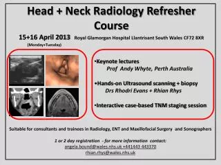
M-1 RADIOLOGY Head and Neck. OBJECTIVES. Skull anatomy Orbit anatomy Sinus anatomy Vascular anatomy Neck anatomy Clinical cases. CASE HISTORIES: Facial pain Trauma to orbit Epistaxsis Allergic reaction Bruit heard in neck Neck mass inferiorly Hoarseness Blurred vision.

E N D
M-1 RADIOLOGY Head and Neck
OBJECTIVES • Skull anatomy • Orbit anatomy • Sinus anatomy • Vascular anatomy • Neck anatomy • Clinical cases
CASE HISTORIES: • Facial pain • Trauma to orbit • Epistaxsis • Allergic reaction • Bruit heard in neck • Neck mass inferiorly • Hoarseness • Blurred vision
SINUSES PA view Nasal Septum Frontal Sinus Maxillary Sinus Ethmoid Sinus Inferior Turbinate Odontoid process Superior orbital fissure 2 7 4 3 1 5 6
AP WATERS VIEW SINUSES 1. Frontal sinus 2. Ethmoid Sinus 3. Nasal Septum (bony) 4. Zygomatical-Frontal Suture 5. Maxillary Sinus 6. Zygoma 7. Zygomatic Arch 8. Mandible 9. Inferior orbital margin 10. Left orbit 1 10 2 4 5 3 6 9 7 8 This view is angled to project the maxillary sinuses free of the petrous ridge.
1. Frontal Sinus 2. Maxillary Sinus 3. Ethmoid Sinus 4. Spenoid Sinus 5. Sella Turcica 6. Occipital Bone 7. Mastoid Air Cells 8. Floor of posterior fossa 9. Anterior arch of C-1 10. Mandible 11.Coronal Suture 11 1 3 5 7 2 4 6 9 8 10 LATERAL SINUS & SKULL
SKULL Townes view 1. Parietal bone 2. Lambdoid suture 3. Foramen magnum 4. Petrous temporal bone 5. Mandible 6. Mastoid air cells 1 1 2 3 4 6 5
1. Lat. & Med. ptyergoid plate • 2. Ethmoid Sinus • 3. Odontoid Process • 4. Sphenoid Sinus • 5. Foramen ovale • 6. Maxillary Sinus • 7. Mastoid air cells • 8. Ant arch of C-1 • 9. Margin of foramen magnum • 10. Ext. auditory canal 6 2 1 5 4 10 8 3 7 9 BASE OF SKULL
CAROTID CANAL JUGULAR FORAMEN CT SKULL BASE
MANDIBULAR CONDYLE MASTOID AIR CELLS CT SKULL BASE
ZYGOMATIC ARCH EXTERNAL AUDITORY CANAL CT SKULL BASE
FORAMEN OVALE FORAMEN SPINOSUM CT SKULL BASE
MALLEUS INCUS MIDDLE EAR OSSICLES CAROTID CANAL CT SKULL BASE
CAROTID CANAL OSSICLES MALLEUS INCUS IAC INTERNAL AUDITORY CANAL CT SKULL BASE
3 • LATERAL NECK • Hard pallate • Soft pallate • Nasopharynx • Oropharynx 1 2 4
AIRWAY • Calcified tracheal cartilage rings • Hyoid bone • Epiglottis • Thyroid cartilage • Cricoid cartilage 3 2 5 4 1 LATERAL VIEW OF NECK
SWALLOWING STUDY 1 2 4 3 Note hyoid bone moves anteriorly and superiorly with swallowing.
ARTERIOGRAM • Internal Carotid Artery • Intracranial Carotid • Maxillary Artery • Occipital Artery • External Carotid Artery • Common Carotid Artery • Facial Artery 2 3 4 7 1 5 6
COLOR DOPPLER ULTRASOUND OF CAROTID ARTERY COMMON CAROTID ARTERY
CT- SINUS AXIAL VIEW 1. Frontal Sinus 1 Scans start superiorly and are shown going inferiorly
CT- SINUS AXIAL VIEW 1. Ethmoid Sinus 2. Sphenoid Sinus 3. Carotid canal 1 2 3
CT- SINUS AXIAL VIEW • Maxillary Sinus • Med. & Lat. Pterygoid plate • Nasopharynx • Nasal septum • Inferior turbinate 1 4 5 2 3
CT- SINUS AXIAL VIEW 1 • Maxillary Sinus • Hard Palate • Mandible • Masseter muscle 2 4 4 3 3
CT- SINUS Coronal sections extending from anterior to posterior 2 • Fronto-nasal suture • Frontal sinus • Nasal bones 1 3
CT- SINUS CORONAL VIEW 1 1. Ethmoid sinus 2. Inferior turbinate 3. Middle turbinate 3 2
CT- SINUS CORONAL VIEW • Maxillary sinus • Nasal Septum 1 2
CT- SINUS CORONAL VIEW 1 • Sphenoid sinus • Hard Palatte 2
CT ORBIT AXIAL SCAN • Retrorbital fat • Medial rectus • Lens • Lateral rectus • Optic nerve 2 4 1 3 5
LT MAXILLARY SINUSES ZYGOMA SCAN LEVEL ZYGOMA SPHENOID SINUS Sections from the skull base extending inferiorly through the neck.
MANDIBULAR CONDYLE LT MAXILLA SCAN LEVEL EXTERNAL AUDITORY MEATUS NASOPHARNYX MASTOIDS
MANDIBLE LT MASSETER MUSCLE MASSETER MUSCLE SCAN LEVEL PTERYGOID MUSCLES PAROTID GLAND
LT SUBMANDIBULAR GLAND SCAN LEVEL EPIGLOTTIS STERNOCLEIOMASTOID MUSCLE SUBCUTANEOUS FAT
LT HYOID BONE SCAN LEVEL VALLECULA PRYIFORM SINUS JUGULAR VEIN JUGULAR VEIN COMMON CAROTID ARTERIES
LT STERNOCLEIDOMASTOID MUSCLE SCAN LEVEL THYROID CARTILAGE VOCAL CORD
LT THYROID CARTILAGE SCAN LEVEL COMMON CAROTID ARTERY CRICOID CARTILAGE JUGULAR VEIN
LT SCAN LEVEL THYROID GLAND CLAVICLE CLAVICLE FAT FAT ESOPHAGUS TRACHEA
CASE HISTORIES: • Facial pain • Trauma to orbit • Epistaxsis • Allergic reaction • Bruit heard in neck • Neck mass inferiorly • Hoarseness • Blurred vision
FACIAL PAIN AND NASAL CONGESTION
Note the opacified left maxillary sinus with fluid layering dependently.
CT SINUS MAXILLARY ETHMOID AXIAL SCAN Opacified maxillary and ethmoid sinuses
CT SINUS CORONAL SCAN Opacified left maxillary sinus. The patient has had surgery on right maxillary sinus to create a nasal antral window to improve drainage.
CT SINUS AXIAL SCAN normal Note the destroyed posterior wall of the left frontal sinus due to bacterial invasion.
ORBITAL FLOOR FRACTURE Arrow points to bone fragment displaced into orbit.
CT FACIAL CORONAL SCAN CT scans redemonstrate fracture and edema at site.
TRIPOD FRACTURE • Zygomatic-frontal suture • Zygomatic arch • Maxillary sinus wall Trauma to the maxilla can fracture at the 3 sites creating a Tripod fracture.
ZYGOMATIC FRACTURE
Here external carotid arteriogram shows abnormal vessels in the nasal cavity due to vascular tumor.
Vascular Tumor Normal Tumor seen is rare but typical for angiofibroma