Download
1 / 41
410 likes | 597 Vues
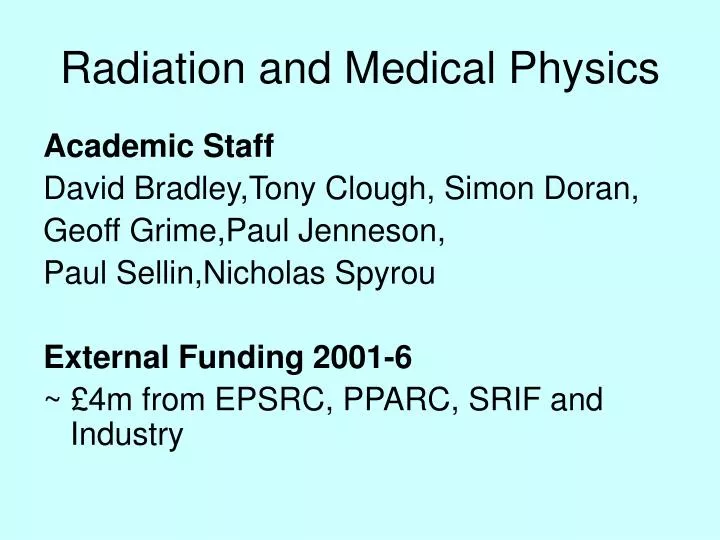
Radiation and Medical Physics. Academic Staff David Bradley,Tony Clough, Simon Doran, Geoff Grime,Paul Jenneson, Paul Sellin,Nicholas Spyrou External Funding 2001-6 ~ £4m from EPSRC, PPARC, SRIF and Industry.

E N D
Radiation and Medical Physics Academic Staff David Bradley,Tony Clough, Simon Doran, Geoff Grime,Paul Jenneson, Paul Sellin,Nicholas Spyrou External Funding 2001-6 ~ £4m from EPSRC, PPARC, SRIF and Industry
The research of the group covers many areas, from radiation detector development to NMR in medicine. Topics to be covered today: • Muon Radiography • 3- Positron tomography • Ion Beam Analysis of Materials
MUON RADIOGRAPHY OF LARGE INDUSTRIAL STRUCTURES Walter Gilboy, Paul Jenneson - Surrey University Stefaan Simons - University of London Steven Stanley, Dominic Rhodes – British Nuclear Fuels
COSMIC RADIATION “It falleth as the gentle rain from heaven” (Apologies to Shakespeare) Sea level muon intensity 10,000 m-2 min-1 It’s free, universally available andVERY GREEN!
Monitoring Blast Furnace liner thickness Blast Furnace • A very large steel containing vessel lined internally with up to 2 metres of carbon heat insulation below 1 metre liquid iron The Problem • The liner gradually wears away and when it reduces below 50 cm the danger of molten iron breakthrough through the base increases. A possible monitoring solution • When negative muons are reduced to rest in matter nuclear absorption of negative muons can occur. • The rate is approximately proportional to Z4 so that absorption in iron (Z=26) is much more likely than absorption in carbon (Z=6). The resulting nuclear excitation gives rise to neutron emission and the greater yield from the higher Z material can be used to distinguish the two materials. • The carbon acts as a neutron moderator and thermalised neutrons diffusing out of the furnace can be detected relatively easily. • The potential of this approach has been investigated with a simple experimental set-up and the results compared to Monte Carlo predictions.
Incident Cosmic ray Muons Wooden pallet Cadmiumsheet Muon induced neutron experimental layout
µs mainly stopping in Pb µs mainly stopping in C Relative Count Rate C moderator too thin
THREE-GAMMA IMAGING IN POSITRON EMISSION TOMOGRAPHY N.M. Spyrou and K. Kacperski Centre for Nuclear and Radiation Physics University of Surrey, U.K.
Conventional PET • Mainly BGO (Bismuth Germanate) scintillation • detectors used: • High stopping power gives: • good efficiency • good spatial resolution • Good timing • Poor energy resolution (12% FWHM @ 511keV at best) 511 keV 511 keV Can we get more out of PET ? Is there more information in the annihilation radiation that could possibly be extracted ? Yes!
511 keV 511 keV 511 keV 511 keV Physics of positron annihilation slow down Positronium formation (30%) Annihilation with free electron (70%) 75% 25% p-Ps t =0.125 ns o-Ps Conversion 99.73% t =142 ns 0.27% 98.7% 1.3% Pick-off 2g 3g 3g 2g Chemical reactions Altogether ~ 0.5% of 3 annihilations
How can the 3g annihilations be useful? Extracting more information • Although 3 events are rare compared 2 events, the information obtained per event is greater than for 2annihilation • It is NOTan alternative but an addition to conventional PET imaging • Additional information gained may lead to reduced administered dose Tumour Hypoxia • The 3 yield is sensitive to the local chemical environment and may be able to confirm the level of oxygenation in tumours
z D r 2 - - - E x x E x x E x x r E E r = + + = 3 3 1 1 2 2 p 0 1 2 1 x - - - h c r r c r r c r r y 1 2 3 E - - - E y y E y y E y y 3 = + + = 3 3 1 1 2 2 p 0 y - - - r c r r c r r c r r 1 2 3 3 - - - E z z E z z E z z = + + = 3 3 1 1 2 2 p 0 z - - - c r r c r r c r r 1 2 3 x + + = 2 E E E 2 m c 1 2 3 e The concept of 3g imaging in PET g 3 photons of energies E , E , E 1 2 3 r r r , , detected in coincidence at 1 2 3 From the law of momentum conservation: 2D or 3D set of nonlinear equations r Solve for The position of annihilation site recovered from a single event!
... ... Coincidence SCA Preamp / Amp Gating New PET scanner design CdZnTe scanner g g 2 3 r ,r ,r ; E , E , E r ,r List mode 1 2 3 1 2 3 1 2 ... ... NO Is E , E , E > E ? reject 1 2 3 min < E max YES NO ? reject E + E + E = 1022keV 1 2 3 YES r solve for conventional YES g ? r Is within the object 3 image PET image Add to image NO reject
Conclusions • 3 annihilation events can be used for imaging in PET • The information conveyed by single 3 event is higher • New type of scanner may give an overall improvement in PET imaging at lower doses • There are only few 3 events • High resolution CZT detectors are required to make 3 imaging possible; scintillation detector energy resolution is insufficient • The quality of the 3 image will be rather poor. Further Research Direction: Can the differences of 3 yields in tissues be used to measure tumour hypoxia?
Ion Beam Analysis at Surrey Accelerator: 2MV Tandetron Ion source: 3He, 4He or protons • External Scanning microbeam • (~ 10 microns spot size) • 3mm diameter scan size Tandetron Magnet Object aperture Quadrupole focussing magnets Computer controlled raster scanner deflection plates Scanning microbeam target chamber LN2 cooled sample stage
Applying External Micro-PIXE Technique to Investigate Chlorine Diffusion In Mortars AS Clough MS Rihawy FE Gauntlett MJ Mulheron
Surrey External Micro-Beamline Beam from Tandetron
Chloride Diffusion from a Mortar Mortar Cylinder A cylindrical sample of a mortar, containing 1% NaCl by weight, is immersed in water. After immersion the sample is sectioned in planes -one parallel to, one perpendicular to the base - to study chlorine diffusion horizontally and vertically. Water
Samples were placed in the external ion beam, raster scanned over an area 3mm diameter then moved in steps of 3mm to allow a series of scans across the section. Characteristic X-rays for elements found in the samples were mapped.
Cl Ca Fe Si K
Horizontal Scan 3 mm
Conclusion Scanning external beam MeV PIXE analysis provides a simple, rapid and low cost method of determining chlorine profiles in mortars at low levels (~ 0.5%) by weight with a spatial resolution of about 20 microns. We intend to extend this study to various kinds of mortars and concrete.
Ion Beam Developments at Surrey Horizontal nanobeam for ion beam writing and ion beam analysis Vertical single-ion beamline for studying interactions with cells - pioneered independently at Columbia University, NY and the Gray Laboratory, Middlesex by Alumni:David Brenner and Melvyn Folkard. External Scanning microbeam (~ 10 microns spot size) Vertical and horizontal nanobeams (<10 nm spot size) under construction Tandetron Magnet Object aperture Quadrupole focussing magnets Computer controlled raster scanner deflection plates Scanning microbeam target chamber LN2 cooled sample stage
Ion microbeam analysis of diffusion in materials Tony Clough, Fern Gauntlett, Salah Rihawy • ION MICROBEAM ANALYSIS TECHNIQUE • APPLICATIONS • FUTURE WORK
Scanning Microbeam Target Chamber Side view Front view of sample stage Particle detectors Cu blocks X-ray detector Sample Scanning zone Focussed 3He or p scanning microbeam LN2 cooled sample stage HPGe detector
NRA (Nuclear Reaction Analysis) Protons detected from the reaction: 3He + d p + Q = 18.4 MeV PIXE (Particle Induced X-ray Emission) Ions can scatter inner electrons from a target atom. As the electrons rearrange, X-rays characteristic of the target element are produced. L X-rays are produced from the transition of electrons from outer shells to the L shell. K X-rays are produced from the transition of electrons from outer shells to the K shell.
Water diffusion into fibre optic pressure sensors • Fibre optic ~ 125 m in diameter soaked in D2O at 210°C and 19 bar for 50 days • 1 cm long sections mounted between copper blocks on sample plate and circular cross-section cleaved at block surface. • Plate mounted in scattering chamber, cooled with LN2 and the fibre cross-section exposed to a scanning 2 MeV 3He beam. The detected protons and X-rays were recorded as a function of beam position.
Copper sample holder Tissue sample Scanned regions appear darker Figure 10: Porcine muscle tissue in copper blocks sample holder, scanned areas visible as discolouration on meat
PIGE - F (drug) RBS- Cu (sample holder)