Download
1 / 64
660 likes | 868 Vues
Chapters 31 and 32 Anatomy and Physiology of the Male and Female. Introduction. Proper functioning of the reproductive system ensures the survival of the species

E N D
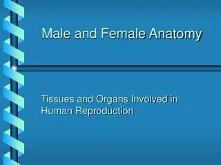
Chapters 31 and 32Anatomy and Physiology of the Male and Female
Introduction • Proper functioning of the reproductive system ensures the survival of the species • Male reproductive system consists of organs whose functions are to produce, transfer, and introduce mature sperm into the female reproductive tract where fertilization can occur
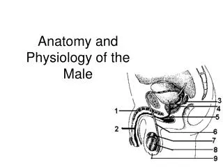
Male Reproductive Organs • Classified as essential organs for production of gametes or accessory organs that support the reproductive process (Figure 31-1, A) • Essential organs—gonads of the male; testes • Accessory organs of reproduction • Genital ducts convey sperm to outside of body; pair of epididymides, paired vasa deferentia, pair of ejaculatory ducts, and the urethra • Accessory glands produce secretions that nourish, transport, and mature sperm; pair of seminal vesicles, the prostate, and pair of bulbourethral glands • Supporting structures—scrotum, penis, and pair of spermatic cords
Anatomy of Male Reproductive System • Perineum—in males, roughly diamond-shaped area between thighs; extends anteriorly from symphysis pubis to coccyx posteriorly; lateral boundary is the ischial tuberosity on either side; divided into the urogenital triangle and the anal triangle (Figure 31-1, B)
Testes • Structure and location • Located within the scrotum • Spermatic cord contains the nerves and blood vessels which supply the testes • Several lobules composed of seminiferous tubules and interstitial cells (of Leydig), separated by septa, encased in fibrous capsule called the tunica albuginea (Figure 31-2) • Seminiferous tubules in testis open into a plexus called rete testis, which is drained by a series of efferent ductules that emerge from top of organ and enter head of epididymis
Testes • Functions 1. Spermatogenesis—formation of mature male gametes (spermatozoa) by seminiferous tubules via meiosis 2. Secretion of the hormone, testosterone, by interstitial cells • Allows development of male characteristics • Promotes growth of skeletal muscle and closure of the growth plate • Small amounts of estrogen are also normal (regulates spermatogenesis, promotes sexual behavior and partner preference) • Table 31-1 summarizes male reproductive hormones
Testes • Sperm development • Spermatogonia – primary spermatocyte – secondary spermatocyte – spermatids – spermatozoa • Capacitation – the final development process sperm undergo while in the vagina • Structure of spermatozoa (Figure 31-7) • head which contains the genetic material • An acromsome contains hydrolytic enzymes to allow sperm to penetrate the egg • Midpiece contains mitochondria to power movement of the tail
Reproductive (Genital) Ducts • Epididymis • Structure and location • Single tightly coiled tube enclosed in fibrous casing (Figure 31-8) • Lies along top and side of each testis • Anatomical divisions include head, body, and tail • Functions • Store sperm for 1-3 weeks • Produce seminal fluid (semen) • Sperm become capable of motility while they are passing through epididymis
Reproductive (Genital) Ducts • Vas deferens (ductus deferens) • Structure and location • Muscular tube; extension of epididymis • Extends through inguinal canal, into abdominal cavity, and over top and down posterior surface of bladder • Enlarged terminal portion called ampulla—joins duct of seminal vesicle • A Vasectomy makes a man sterile because it interrupts the route from the epididymis • Function • One of excretory ducts for seminal fluid • Connects epididymis with ejaculatory duct • Ejaculatory duct • Formed by union of vas deferens with duct from seminal vesicle • Passes through prostate gland, terminating in urethra • Urethra
Accessory Glands • Seminal vesicles • Empty into ejaculatory duct • Produce about 60% of semen which contains fructose • High pH to help neutralize the environment of the urethra and vagina • Prostate gland • Produces about 30% of semen • Thin, milky secretion, slightly acidic pH, citrate nutrients for sperm • Bulbourethral glands • Contribute about 5% to semen • Mucous secretion. Just before ejaculation • Helps neutralize pH of female vagina
Supporting Structures • Scrotum • Skin-covered pouch suspended from perineal region • Divided into two compartments • Contains testis, epididymis, and lower part of a spermatic cord • Line by the tunica vaginalis • Dartos and cremaster muscles elevate the scrotal pouch • Sexual arousal, cold temperatures and threat of injury cause contraction
Penis • Functions: urination, serves as vehicle for sperm to move into vagina • Three columns of erectile tissue surround the penis • Corpora cavernosa (2). • Corpus spongiosum (1) surrounding spongy urethra Forms the bulb • These tissues fill with blood to create an erection • Glans penis: enlargement of corpus spongiosum • Prepuce or foreskin covers glans penis. Circumcision: surgical removal • External urethra orifice • Scrotum and the penis make up the genitals
Supporting Structures • Spermatic cords (internal) • Fibrous cylinders located in inguinal canals • Encloses the vas deferens, blood vessels, lymphatics, and nerves
Composition and Course of Seminal Fluid • Consists of secretions from testes, epididymides, seminal vesicles, prostate, and bulbourethral glands • Each milliliter contains millions of sperm • Passes from testes through epididymis, vas deferens, ejaculatory duct, and urethra
Male Fertility • Relates to many factors—number of sperm; size, shape, and motility • Fertile sperm are uniform in size and shape and are highly motile • Infertility may be caused by antibodies some men make against their own sperm, “immune infertility”
Cycle of Life: Male Reproductive System • Reproductive functions begin at time of puberty • Development of organs begins before birth; immature testes descend into scrotum before or shortly after birth • Puberty—high levels of hormones stimulate final stages of development • System operates to permit reproduction until advanced old age • Late adulthood—gradual decline in hormone production may decrease sexual appetite and fertility
Descent of Testes • Descent • Pass from abdominal cavity through inguinal canal to scrotum • Pass through inguinal rings (bilateral oblique passageways in the anterior abdominal wall). Openings in aponeuroses of transversus abdominis, internal and external oblique. Weak part in abdominal wall. Herniation. • Cryptorchidism • Failure of one or both of testes to descend into scrotum • Prevents normal sperm development
Anatomy of Female Reproductive System • Female reproductive organs • Ovaries • Uterine tubes • Uterus • Vagina • External genital organs • Mammary glands
Overview of the Female Reproductive System • Function of the female reproductive system • The function of the female reproductive system is to produce offspring and thereby ensure continuity of the genetic code • It produces eggs, or female gametes, which each may unite with a male gamete to form the first cell of an offspring • It also can provide nutrition and protection to the offspring for up to several years after conception
Overview of the Female Reproductive System • Structural plan of the female reproductive system • Reproductive organs are classified as essential or accessory (Figure 32-1) • Essential organs—gonads are the paired ovaries; gametes are ova produced by the ovaries—the ovaries are also internal genitals • Accessory organs • Internal genitals—uterine tubes, uterus, and vagina: ducts or duct structures that extend from the ovaries to the exterior • External genitals—the vulva • Additional sex glands such as the mammary glands
Overview of the Female Reproductive System • Perineum • The perineum is the skin-covered region between the vaginal orifice and the rectum • Divided into the urogenital triangle and the anal triangle • This area may be torn during childbirth • An episiotomy, a surgical incision in the perineum, may be made to avoid tearing
Ovaries • Location of the ovaries • Nodular glands located on each side of the uterus, below and behind the uterine tubes (Figure 32-2) • Supported by the broad and ovarian ligaments • Ectopic pregnancy—development of the fetus in a place other than the uterus • Microscopic structure of the ovaries • Surface of the ovaries is covered by the germinal epithelium • Ovarian follicles contain the developing female sex cells (Figure 32-3) • Ovum—an oocyte released from the ovary
Ovary Histology • Outermost covering: ovarian (germinal) epithelium: visceral peritoneum • Tunica albuginea: capsule of dense fibrous connective tissue • Ovary itself • Cortex: outer, dense. Contains follicles with oocytes • Medulla: inner, looser. Contains blood vessels, nerves, lymphatic vessels • Stroma: connective tissue of the ovary
Maturation and Fertilization of Oocyte • Oogenesis is the production of a secondary oocyte in ovaries • Oogonia are cells from which oocytes develop. The oogonia divide by mitosis to produce other oogonia and primary oocytes. • Five million oocytes produced by the 4th month of prenatal life. About 2 million begin first meiotic division but stop at prophase. All remain at this state until puberty. • Primary oocytes are surrounded by granulosa cells (which produce estrogen) and called a primordial follicle • Primordial follicle becomes a primary follicle when oocyte and granular cells enlarge • Primary follicle becomes secondary follicle and enlarges to form mature or Graafian follicle • Usually only one is ovulated, others degenerate
Ovulation, Fertilization, Follicle Fate • Ovulation: release of a secondary oocyte from an ovary. Unlike spermatogenesis, division of cytoplasm during meiosis is uneven and polar bodies are very small, oocyte very large • Graafian follicle become corpus luteum (which secretes progesterone) • Fertilization: begins when a sperm cell binds the plasma membrane of secondary oocytes and penetrates into cytoplasm. • Secondary oocyte completes meiosis II forming one polar body. Fertilized egg now a zygote • Fate of corpus luteum • If fertilization occurs, corpus luteum persists • If no fertilization, becomes corpus albicans (white scar tissue)
Ovaries • Functions of the ovaries • Ovaries produce ova—the female gametes • Oogenesis—process that results in formation of a mature egg (Figure 32-4) • The ovaries are endocrine organs that secrete the female sex hormones (estrogens and progesterone) to help regulate the reproductive cycle
Uterus • Parts: body, cervix, and fundus • Ligaments: broad, round, uterosacral • Broad: suspends the uterus • Round: from uterus through inguinal canals to labia majora • Uterosacral: attach lateral wall of uterus to sacrum • Composed of 3 layers • Perimetrium: Outer serous membrane • Myometrium: Middle layer of smooth muscle used to expel the fetus • Endometrium: Mucous membrane • Functional layer: innermost. Replaced monthly during menstrual cycle • Basal layer: deepest • Mucus gland secretions normally act as a barrier to sperm but become more slippery during ovulation to help sperm movement
Uterus • Cavities of uterus • The body cavity’s apex constitutes the internal os and opens into the cervical canal, which is constricted at its lower end and forms the external os that opens into the vagina • The blood to the uterus is supplied by uterine arteries
Uterus • Location of the uterus • Located in pelvic cavity between urinary bladder and rectum (Figure 32-1) • Position of uterus (Figure 32-5) is altered by age, pregnancy, and distention of related pelvic viscera • Descends, between birth and puberty, from the lower abdomen to the true pelvis • Begins to decrease in size at menopause
Uterus • Position of the uterus • Body lies flexed over the bladder • Cervix points downward and backward, joining the vagina at a right angle • Several ligaments hold the uterus in place but allow some movement
Uterus • Functions of the uterus (womb) • Part of reproductive tract, permits sperm to ascend toward uterine tubes • If conception occurs, an offspring develops in the uterus • Implants in the endometrial lining • Embryo is supplied with nutrients by endometrial glands until the production of the placenta • Placenta is an organ that permits exchange of materials between mother’s blood and fetal blood but keeps the two circulations separate • Myometrial contractions occur during labor and help push the offspring out of mother’s body • If conception does not occur, outer layers of endometrium are shed during menstruation • Menstruation is a cyclical event that allows the endometrium to renew itself
Uterine (Fallopian) Tubes • Open directly into peritoneal cavity to receive oocyte from ovary • Transport oocyte or zygote from ovary to uterus • Consists of the isthmus, ampulla and infundibulum • Fimbriae: long thin processes that extend from the infundibulum. Inner surface is ciliated. Infundibulum is open to peritoneal cavity • Ampulla: widest part, where fertilization occurs • Three layers: outer serosa, middle muscular layer and inner mucosa. Mucosa: simple ciliated columnar epithelium with longitudinal folds • Provides nutrients for oocyte/embryonic mass • Cilia moves fluid and oocyte/embryonic mass through the tube toward uterus
Vagina • Vagina is a tubular organ located between the rectum, the urethra, and the bladder • Structure of the vagina • Collapsible tube capable of distention, composed of smooth muscle, and lined with mucous membrane arranged in rugae • Anterior wall is shorter than posterior wall because cervix protrudes into its uppermost portion • Hymen—a mucous membrane that typically forms a border around the vagina in young premenstrual girls
Vagina • Functions of the vagina • Receptacle for semen to pool in the anterior and posterior fornix • Lower portion of the birth canal • Transports tissue and blood shed during menstruation to the exterior
Female External Genitalia • Vulva or pudendum or external female genitalia • Vestibule: space • Labia minora: form borders on sides • Clitoris: erectile structure • Labia majora: unite to form mons pubis • Vaginal orifice • External urinary meatus
Female External Genitalia • Within vestibule, glands that produce fluid to moisten • Greater vestibular gland • Lesser vestibular glands • Labia majora: rounded folds of skin on either side of labia minora. Conceal contents of pudenda • Medial surfaces covered with numerous sebaceous and sweat glands • Mons pubis: anteriorly labia majora merge in an elevated area over the symphysis pubis