Download
1 / 2
20 likes | 146 Vues
Understanding rotavirus VP6 nanotube internalization into cells: Towards a recombinant vaccine Journal: Archives of Virology Mabel Rodríguez a , Christopher Wood b , Rosana Sanchez-López c , Ricardo M. Castro-Acosta a , Octavio T. Ramírez a and Laura A. Palomares a *

E N D
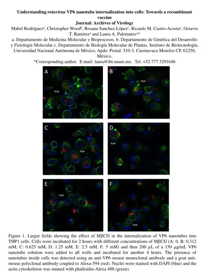
Understanding rotavirus VP6 nanotube internalization into cells: Towards a recombinant vaccine Journal: Archives of Virology Mabel Rodrígueza, Christopher Woodb, Rosana Sanchez-Lópezc, Ricardo M. Castro-Acostaa, Octavio T. Ramíreza and Laura A. Palomaresa* a. Departamento de Medicina Molecular y Bioprocesos. b. Departamento de Genética del Desarrollo y Fisiología Molecular c. Departamento de Biología Molecular de Plantas. Instituto de Biotecnología, Universidad Nacional Autónoma de México, Apdo. Postal. 510-3, Cuernavaca Morelos CP. 62250, México. *Corresponding author. E-mail: laura@ibt.unam.mx. Tel. +52 777 3291646 Figure 1. Larger fields showing the effect of MCD in the internalization of VP6 nanotubes into THP1 cells. Cells were incubated for 2 hours with different concentrations of MCD (A: 0, B: 0.312 mM; C: 0.625 mM, D: 1.25 mM, E: 2.5 mM, F: 5 mM) and then 200 µL of a 150 g/mL VP6 nanotube solution were added to all wells and incubated for another 4 hours. The presence of nanotubes inside cells was detected using an anti-VP6 mouse monoclonal antibody and a goat anti-mouse polyclonal antibody coupled to Alexa-594 (red). Nuclei were stained with DAPI (blue) and the actin cytoskeleton was stained with phalloidin-Alexa 488 (green).
Figure 2. Effect of MCD in the internalization of VP6 nanotubes into J774 cells. Cells were preincubated 2h with different concentrations of MCD (A: 0, B: 0.625mM, C: 1.25mM, D: 2.5mM, E: 5mM) and then 150g/mL of VP6 nanotubes were added and incubated for another 4 hours. The VP6 internalized into cells was detected using fluorescence microscopy with an anti-VP6 mouse monoclonal antibody and a goat anti-mouse polyclonal antibody coupled to Alexa-594 (red) and nuclei were stained with DAPI (blue). The right panels show the green signal corresponding to the actin cytoskeleton stained with phalloidin coupled to Alexa488.