Download
1 / 41
420 likes | 634 Vues
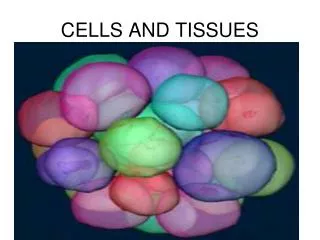
Dr. Hassan Shaibah. Cells: The Living Units. Midbrain@gmail.com Ext.4011. Introduction to Cells. Cells – the smallest living units in our bodies Organelles – “little organs” – carry on essential functions of cells Enzymes – direct chemical reactions in cells

E N D
Dr. Hassan Shaibah Cells: The Living Units Midbrain@gmail.comExt.4011
Introduction to Cells • Cells – the smallest living units in our bodies • Organelles – “little organs” – carry on essential functions of cells • Enzymes – direct chemical reactions in cells • Metabolism – the sum of all chemical reactions in the cell
Introduction to Cells • Cells have three main components • Plasma membrane • Cytoplasm • Nucleus
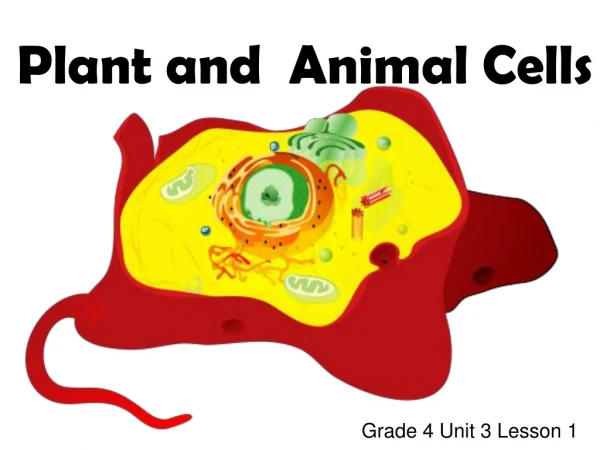
Structure of a Generalized Cell Figure 2.1
The Plasma Membrane • Plasma membrane defines the extent of the cell • Structure of membrane • Fluid mosaic model (lipid bilayer) • Types of membrane proteins • Integral proteins – firmly imbedded in, or attached to lipid bilayer • Peripheral proteins – attach to membrane surface
The Plasma Membrane Figure 2.2a
The Plasma Membrane • Functions – relate to location at the interface of cell’s exterior and interior • Provides barrier against substances outside the cell • Some plasma membranes act as receptors
The Plasma Membrane • Determines which substances enter or leave the cell • Membrane is selectively permeable • Diffusion – molecules move from a region where they are more concentrated to an area where they are less concentrated • Osmosis – the diffusion of water across a membrane
Vesicular or Bulk Transport • Exocytosis – mechanism that moves substances out of the cell • Endocytosis – mechanism by which particles enter cells • Phagocytosis – “cell eating” • Pinocytosis – “cell drinking”
Exocytosis Figure 2.3a
Phagocytosis and Pinocytosis Figure 2.4a, b
Endocytosis • Receptor-mediated endocytosis • Plasma proteins bind to certain molecules • Invaginates and forms a coated pit • Pinches off to become a coated vesicle
Receptor-Mediated Endocytosis Figure 2.5
The Cytoplasm • Cytoplasm – lies internal to plasma membrane • Consists of cytosol, organelles, and inclusions • Cytosol (cytoplasmic matrix) • Jelly-like fluid in which other cellular elements are suspended • Consists of water, ions, and enzymes
Cytoplasmic Organelles • Mitochondria – generate most of the cell’s energy • Most complex organelle • Ribosomes – constructed of proteins and ribosomal RNA • Site of protein synthesis
Cytoplasmic Organelles • Endoplasmic reticulum – “network within the cytoplasm” • Rough ER – ribosomes stud the external surfaces • Smooth ER – consists of tubules in a branching network • No ribosomes are attached; therefore no protein synthesis
Assembly of Proteins at the Rough Endoplasmic Reticulum Figure 2.10
Cytoplasmic Organelles • Golgi apparatus – a stack of three to ten disk-shaped envelopes • Sorts products of rough ER and sends them to proper destination
Role of the Golgi Apparatus in Packaging Products of Rough ER Figure 2.12
Cytoplasmic Organelles • Lysosomes – membrane-walled sacs containing digestive enzymes • Digest unwanted substances • Peroxisomes – membrane-walled sacs of oxidase enzymes • Enzymes neutralize free radicals and break down poisons • Break down long chains of fatty acids • Are numerous in the liver and kidneys
Cytoplasmic Organelles • Cytoskeleton – “cell skeleton” – an elaborate network of rods • Contains three types of rods • Microtubules – cylindrical structures made of proteins • Microfilaments – filaments of contractile protein actin • Intermediate filaments – protein fibers
The Cytoskeleton Figure 2.14
Cytoplasmic Organelles • Centrosomes and centrioles • Centrosome – a spherical structure in the cytoplasm • Composed of centrosome matrix and centrioles • Centrioles – paired cylindrical bodies • Consists of 27 short microtubules • Act in forming cilia
Cytoplasmic Organelles • Vaults – barrel-shaped protein structures (discovered in the late 1980s) • Function unknown • May shuttle large molecules from nucleus to cytoplasm
Cytoplasmic Inclusions • Temporary structures – not present in all cell types • May consist of pigments, crystals of protein, and food stores • Lipid droplets – found in liver cell and fat cells • Glycosomes – store sugar in the form of glycogen
The Nucleus • The nucleus – “central core” or “kernel” – control center of cell • DNA directs the cell’s activities • Nucleus is approximate 5µm in diameter Figure 2.17a
The Nucleus • Nuclear envelope – two parallel membranes separated by fluid-filled space • Chromatin – composed of DNA and histone proteins • Condensed chromatin – contains tightly coiled strands of DNA • Extended chromatin – contains uncoiled strands of DNA • DNA's genetic code is copied onto mRNA (transcription)
The Nucleus • Chromosomes – highest level of organization of chromatin • Contains a long molecule of DNA
The Nucleus • Nucleolus – “little nucleus” – in the center of the nucleus • Contains parts of several chromosomes • Site of ribosome subunit manufacture
Cellular Diversity • Specialized functions of cells relates to: • Shape of cell • Arrangement of organelles
Cellular Diversity • Cells that connect body parts or cover organs • Fibroblast – makes and secretes protein component of fibers • Erythrocyte – concave shape provides surface area for uptake of the respiratory gases • Epithelial cell – hexagonal shape allows maximum number of epithelial cells to pack together
Cells that Connect Body Parts or Cover Organs Figure 2.22(1)
Cellular Diversity • Cells that move organs and body parts • Skeletal and smooth muscle cells • Elongated and filled with actin and myosin • Contract forcefully
Cells that Move Organs and Body Parts Figure 2.22(2)
Cellular Diversity • Cells that store nutrients • Fat cell – shape is produced by large fat droplet in its cytoplasm • Cells that fight disease • Macrophage – moves through tissue to reach infection sites
Cells that Store Nutrients and Cells that Fight Disease Figure 2.22(3), (4)
Cellular Diversity • Cells that gather information • Neuron – has long processes for receiving and transmitting messages Figure 2.22(5)
Cellular Diversity • Cells of reproduction • Oocyte (female) – largest cell in the body • Contains many copies of organelles for distribution to daughter cells • Sperm (male) – possesses long tail for swimming to the egg for fertilization Figure 2.22(6)
Developmental Aspects of Cells • Youth – begin as a fertilized egg • Cells in embryo • Exposed to chemical signals • Chemicals channel cells into specific pathways of development • Cell specialization leads to structural variation of cell types
Developmental Aspects of Cells • Aging – a complex process caused by a variety of factors • Free radical theory • Damage from byproducts of cellular metabolism • Radicals build up and damage essential molecules of cells • Mitochondrial theory – a decrease in production of energy by mitochondria weakens and ages our cells
Developmental Aspects of Cells • Genetic theory – proposes that aging is programmed by genes


![[virtual] cells](https://cdn1.slideserve.com/3553683/slide1-dt.jpg)