Download
1 / 33
330 likes | 519 Vues
TREATMENT AFTER BRAIN INJURY. Teresa Such-Neibar, DO Primary Children’s Medical Center TBI Advisory Committee. Acquired Brain Injury .

E N D
TREATMENT AFTER BRAIN INJURY Teresa Such-Neibar, DO Primary Children’s Medical Center TBI Advisory Committee
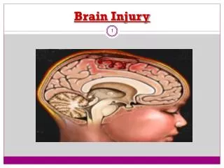
Acquired Brain Injury • An acquired brain injury is an injury to the brain which is not hereditary, congenital, degenerative, or induced by birth trauma. An acquired brain injury is an injury to the brain that has occurred after birth. BIAA website
Neurologic Disorders Commonly Associated with Cognitive Impairment • Traumatic Brain Injury (TBI) • Anoxia/Hypoxia (loss or decrease of oxygen) • Stroke • Infectious Disease (Meningitis) • Demyelinating Disease (MS) • Intracranial Tumors • Toxic Exposure (Carbon Monoxide Poisoning)
Definition - Traumatic Brain Injury • Traumatic brain injury (TBI) is damage to the brain caused by an external physical force. The damage may occur from the movement of the brain within the skull or from penetration of an object into the skull, contacting the brain directly. TBI can result in cognitive dysfunction, physical impairment, or psychological disturbance. BIAA Website
Background • 2 million Americans per year sustain a brain injury. • 200,000 people either die or have severe disability secondary to TBI each year. • The outcome for people with severe brain injuries remains poor with 50% suffering death or severe disability.
Etiology: Common causes of traumatic brain injury include: Falls Motor vehicle accidents Motorcycle accidents Pedestrian-automobile accidents Gunshot wounds – 30% of intentional injuries Shaken baby syndrome Accidents in recreational sports (football, boxing, skiing, etc.) – 1%
Mechanism of Injury • Bruising (bleeding) - Blood vessels can tear when the brain is injured. When this occurs, the blood pools within the brain and begins to press on sensitive brain tissue. The brain tissue will die off and critical parts of the brain may stop functioning. (Hematoma)
Mechanism of Injury • Tearing - Tiny tears can occur when the brain is injured. These tears are usually microscopic and cannot be observed with a CT scan or MRI. (Diffuse Axonal Injury)
Mechanism of Injury • Swelling - Swelling occurs when the body realizes that the brain has been injured. Extra help is sent to help it heal, but because there is very little room within the skull, pressure begins to build up; this can damage parts of the brain. Critical areas within brain may stop functioning. (Edema)
Mechanism of Injury • Primary mechanisms are usually focal in nature such as : • Hematomas
Mechanism of Injury • Contusions
Mechanism of Injury • Penetrating Injuries
Pathophysiology • Acceleration/ deceleration forces • Diffuse Axonal Injury results from stretching and distortion of axons and myelin sheaths.
Pathophysiology • Retraction balls correspond to disruption of axonal transport. • Total axonal disruption is irreversible.
Injury to the Brain • Focal damage is localized and occurs at the point where an object penetrates the brain or where the impact is the greatest. • Diffuse damage occurs when the brain hits the skull during a closed head injury, and it often results in diffuse axonal injury - a tearing of the nerve cells. • The brain stem, as well as the frontal and temporal lobes, are more susceptible to damage because they are located near bony protrusions in the skull.
Secondary Injury Ischemic Brain Injury (decreased oxygen) • Hypoxic insult Edema (increased swelling) • Intracranial Hypertension • Herniation • Hydrocephalus
Glasgow Coma Scale • Eye Opening:Spontaneous E4 To verbal command 3 To pain 2 No response 1 • Best Motor Response: Follows simple commands M6 Localizes pain 5 Localized withdrawal to pain 4 Flexion (decorticate) posturing, pain 3 Extensor (decerebrate) posturing, pain 2 No response 1 • Best Verbal Response: Oriented and converses V5 Disoriented and converses 4 Inappropriate words 3 Incomprehensible sounds 2 No response 1
Glasgow Outcome ScaleJennett, Bond; 1975 1. Death 2. Persistent vegetative state 3. Severe disability (dependent) 4. Moderate disability (independent but disabled) 5. Good recovery (but may not be fully restored)
Look at the Big Picture • Medication is only one factor to consider. • Evaluate the medical history, pain history, previous diagnoses, family history. • Evaluate the environment, ie. the amount of stimulation, present medications . . . are the non-medical options maximized? • Medications should be used as an adjunct therapy and not as the only therapy.
Medication Eval • What are you treating? • Are the non-pharmacological options maximized? • Medications should be used as an adjunct therapy and not as the only therapy option.
Behaviors That May Need Treatment • Agitation • Aggression • Attention/Concentration • Depression/Emotional Lability • Seizures • Sleep
Agitation • Evaluate the Etiology • Overstimulation – Environmental - not punitive • Antidepressants – Tricyclic vs SSRI’s • Beta blockers – antihypertensives • Stimulants, Strattera if d/t frustration • Avoid Benzodiazepines d/t cognitive slowing • Clonidine – monitor blood pressure
Aggression • Attempt to elicit the cause • Lithium for individuals with Bipolar tendencies. • Anticonvulsant medications are the treatment of choice for patients with outbursts of rage and abnormal EEG findings. • Consider a antidepressant prior to the antipsychotics • The Atypical Antipsychotics – olanzapine, risperidone, clonazipine, may help. • Stimulants
Attention/Concentration • Stimulants/Strattera • Tricyclic Antidepressants • Amantadine • SSRI’s • Atypical Antidepressants • Provigil for fatigue
Depression/Emotional Lability • SSRI’s – paroxetine (paxil), sertraline (zoloft), fluoxetine(prozac), citalopram(celexa), escitalopram(lexapro) • Tricyclic Antidepressants – imipramine (tofranil) • Other antidepressants – trazodone, bupropion (wellbutrin), venlafaxine (effexor), nafaxodone (serzone), mirtazapline (remeron)
Seizures • Tegretol – can be used for mood stability • Depokote • Lamictal • Trileptal
Disrupted Sleep • Melatonin – helps regulate sleep cycle – can add Benadryl • Trazodone – difficulty staying asleep • Imipramine – if agitation is also a problem • Ambien – if it is difficulty getting to sleep • Celexa – for sleep and depression or emotional lability • Paxil – if depression and anxiety are issues
General Guidelines • Limit polypharmacy • Use medications that have more than one role/function • Monitor side effects • Re-evaluate frequently
Thank You Questions? Teresa.Such-Neibar@imail.org