Download
1 / 30
330 likes | 563 Vues
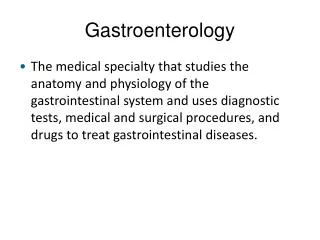
Gastroenterology& hepatology: Introduction. GI Diseases:. Major cause of morbidity & mortality. 10% of GP consultations are for indigestion. ¼ of GP consultations for diarrhea. Infective diarrhea is a major cause of ill heath & death in developing countries.

E N D
GI Diseases: • Major cause of morbidity & mortality. • 10% of GP consultations are for indigestion. • ¼ of GP consultations for diarrhea. • Infective diarrhea is a major cause of ill heath & death in developing countries. • GIT is one of the most common sites for cancer. • Major advances had occurred in the field of GE; • PUD proved to be an infective condition due to HP & Nobel prize had been given recently to its discoverer, Marshal. • Molecular events in the CRC development had been discovered & from this effort became successful in its prevention by NSAIDs. • GIT endoscopy made diagnosis of GIT diseases very easy. • Therapeutic endoscopy made it possible to replace surgery for many GIT conditions as GI bleeding, bilairy stone removal & stenting, palliative cancer stenting, polyp removal, PEG & endoscopic mucosal resection.
GI symptoms: • Dysphagia: difficult swallowing • Odynophagia: painful swallowing. • Aphagia: can not swallow. • Heart burn. • Non cardiac chest pain. • Regurgitation. • Aerophagia: eructation. • Hematemesis. • Melena. • Hematochesia: fresh bleeding per rectum. • Dyspepsia: abnormal digestion. • Anorexia. • Flatulence. • Alteration in bowel habits. • Bleeding per rectum. • Abd pain.
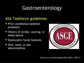
GI Diseases: Investigations • Tests of structure. • Tests of infection. • Tests of function.
1.Tests of structure: Imaging • Plain radiograph: • Show gas within bowel for diagnosis of Int obst if there are dialated loops or fluid levels in the erect position. • Soft tissue of the liver, spleen& kidneys & calcifications in these organs, pancrease,blood vessels, LNs,calculi. • Chest XR in erect position show air under diaphragm in perforated viscus.
Prone Supine LBO
Tests of structure: Imaging • Contrast studies: • Barium & double-cnotrast barium using air with barium, will show filling defects, strictures, erosions & ulcers & even motlity disorders if under fluroscopy.
Tests of structure: Imaging • Contrast studies: uses & limitations.
Tests of structure: Imaging • U/S,CT,MRI:Increasingly used for abd diseases, noninvasive & offer detailed images of abd contents.
Tests of structure: endoscopy • Endoscpy: UGI, LGI, Enteroscopy, ERCP,EUS , Double balloon endoscopy, capsule video endoscopy. • Increasingly used for abd diseases, noninvasive & offer detailed images of abd contents.
Tests of structure: endoscopy • Endoscpy: UGI: Indications: • Dyspepsia sp > 45. • Abd pain. • Atypical chest pain. • Dysphagia • Vomiting. • Wt loss. • Acute or chronic GIB. • Suspicious Ba meal.
Tests of structure: endoscopy • Endoscpy UGI: contraindications: • Severe shock. • Recnet AMI, Unstable angina or arrhythmia. • Severe resp dis. • Atlanto axial sublaxation. • Suspected perforated viscus. • These may be relative in experienced hands. • Endoscpy UGI: Complications: • Cardiorespiratory depression due to sedation. • Aspiration pneumonia. • Perforation. • Bleeding. • SBE( needs prophylaxis in those at risk).
Tests of structure: endoscopy • Colonoscopy: indications: • Suspected IBD. • Altered bowel habits. • Rectal bleeding or anemia. • Suspected abn Ba enema. • CRC screening. • Therapeutic procedure. • C/Is: • Severe shock, Recent AMI, unstable angina & arrhythmias,Severe resp disease,Suspected perforation,severe active UC. • Complications: • As for upper GI endoscopy.
ERCP – DILATED COMMON BILE DUCT DUE TO MULTIPLE GALLSTONES
Tests of structure: Biopsy • Obtained through endoscpy or percutanously & sent for histopath exam. • Reasons for biopsy or cytological exams: • Brash cytology of suspected malignant lesions. • Histological assessment of mucosal abns. • Diagnosis of infections( candida, HP,Giardia). • Measure enzymes as disacharidases. • Analysis of genetic mutations as oncogenes , tumor suppressor genes.
2.Tests of infection: Bacterial cultures • For identifying causes of diarrhea sp if acute or bloody. • Causes of infective diarrhea: • Viruses: Rota, adeno, entero, requires EM or viral cultures. • Bacteria: Campylo jej, EC,Salmonella,clostridium difficile( ned toxine isolation). • Protozoa: Giardia,ameba, cryptosporidium & moicrospora.
Tests of infection: serology • Sp for HP, Salmonella, hydatid liver & ameba.
Tests of infection: radioactive breath tests • For diagnosis for HP & small intestinal bacterial overgrowth.
3.Tests of function: blood tests for malabsorption • S.B12, folic acid, iron, Ca, alumin, phosphate,stool fat, endoscopic DU biopsy. • Tests for diagnosing fat, lactose,bile acids malabsorption. • Tests of pancreatic exocrine function
Tests of function: GIT motility • Esophageal motlity: • Eso manometry with 24 hour Ph monitoring: for diagnosing refractory GERD, Achalasia & noncardiac chest pain. • Gastric motility: • Assessment of gastric emptying in patients with gastropariesis, is best evaluated by radioisotope studies by a test meal of solid & liquid labeled with different radioisotopes. • Small intestine transit: • Difficult & rarely needed. • Ba follow through can measure SI transit to reach TI( 90 min). • Orocecal transit is measured by lactulose-hydrogen breathtest. • Colonic & anorectal motility: • Assessed by anorectal manometry, EP tests, defecating proctography. • Plain Abd XR taken on day 5 after ingestion of different-shaped inert plastic pellets on day 1-3 gives estimate of whole gut transit time. • Help to diagnose chronic idiopathic constipation from mechanical or obstructed defecation.
Tests of function: Radioisotope tests • Gastric emptying study: by Tc. • Urea breath test: by radioactive Carbon: for HP diagnosis as HP have urease which split radioactive urea into amonia & CO2 measured in the breath. • Meckels sacn: TC. • Labeled RBC scan FOR DETECTING BLEEDING. • Labled WBC scan: for detecting infection or inflammation. • Triolin test: C14 labeled trioliln: for fat malabsorption. • Labelled albumin: to detect protein-losing enteropathy.