Download

1 / 31
310 likes | 501 Vues
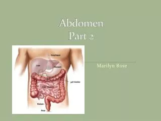
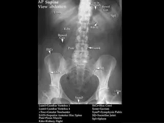
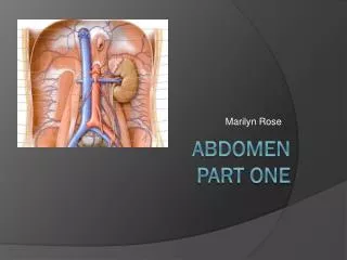
Abdomen-Part 3. Marilyn Rose. Anatomy of a Kidney. Urinary System. Kidneys, ureters, bladder, urethra Kidneys- retroperitoneal, bean-shaped Oblique orientation, paravertebral gutters along posterior abdominal wall One on each side of spine at the level of T12 – L4

E N D
Abdomen-Part 3 Marilyn Rose
Urinary System • Kidneys, ureters, bladder, urethra • Kidneys- retroperitoneal, bean-shaped • Oblique orientation, paravertebral gutters along posterior abdominal wall • One on each side of spine at the • level of T12 – L4 • Surrounded by perirenal fat, and Gerota’s fascia • Composed of • outer cortex- functional subunit- nephrons- filter urine • inner renal medulla- pyramids, Loops of Henle
Kidney on CT Mass on CT recon of Horseshoe Kidney
Adrenal Glands • Retroperitoneal, paired, suprarenal, y shaped • Rt adrenal- posterior to IVC, medial to Rt hepatic lobe, lateral to Rtcrus of diaphragm. • Lt adrenal- in “triangle” of the aorta, pancreatic tail, and Lt kidney- borders Lt crus of diaphragm • Outer cortex- produce steroids • Corticosteroids • glucocorticoids, mineralocorticoids and androgens • Inner medulla • Hormones • Epinephrine and norepinephrine- fight or flight
Adrenal Gland- Abnormal Neuroblastoma Of Lt adrenal gland
Abnormal Adrenal Adrenal Hemorrhage Neuroblastoma
GI System • Stomach- food reservoir and early digestion • Located under Lt dome of diaphragm • Superior portion- joins esophagus at cardiac orifice (cardiac sphincter) • Boder- lesser and greater curvature • Inferior portion- pyloric antrum -> duodenum • Anterior surface- contact with diaphragm, anterio abdominal wall and Lt lobe of liver • Lining of stomach = rugae • Gastric juices= mucus, hydrochloric acid, intrinsic factor and pepsinogen and lipase • Very vascular organ
Stomach on CT CT scan of chest & upper abdomen (coronal section); herniation of stomach & splenic flexure of colon, along with collapse of lung and mediastinal shifting
Intestines • Small bowel • between pylorus and ileocecal valve • 6-7 meters in length • Duodenum • Pylorus- head of panc- retroperitoneal • 4 portions • First- superior-duodenal bulb • Second- descend- ampulla of Vater • Third- horizontal- L3- ant to SVC, AO • Fourth- ascending- Lt of AO at L2 meets with Jejunum • jejunum– duodenojejunal flexure • ligament of Treitz- suspensory lig-around celiac axis • Entry of small bowel into peritoneal cavity • Lt upper abdomen/ umbilical region- absorption occurs- folds?? • Ileum • Longest portion, RL abdomen- terminate at ileocecal valve- CECUM • Often this is the site of intussusception / inflammation and WHAT?
Bowel Issues! Intussesseption hernia
Large Intestine • Inferior to stomach and liver • Larger diameter, haustra and bands called taenia coli • The appendix attaches to posteromedial surface of cecum • Ascending- • retroperitoneal, cecum to liver- hepatic flexure • Transverse • Peritoneal, horizontal, toward spleen, splenic flexure • Descending • Retroperitoneal, Lt lat abd to sigmoid • Sigmoid
Lymph Nodes • Chains along branches of arteries of intestine and AO • Small, oblong, soft and difficult to visualize –unless ABNORMAL • Enlarged= greater than 1 cm in short axis and 2 cm in long axis. • Abdominoaortic nodal groups- surround AO/IVC • Visceral –drain adjacent organs • Lymph drains from abdominal cavity into lumbar trunk, and intestinal in to intestinal trunk and both trunks join the thoracic duct and then the venous system
Lymph Nodes PTLD????
Muscles • Diaphragm • Quadratuslumborum • Lg portion of posterior abdominal wall • Iliac crest- inferior 12th rib • Psoas • Lateral surpaces of lumbar vertebrae • Insert into greater trochanter of femur • Rectus abdominis • Anterior surface of abd/pelvis • Linea alba • Xiphoid- symphysis pubis- midline and interlacing