Download
1 / 72
840 likes | 1.85k Vues
Local Anesthesia Techniques Part 1. Dr. Rahaf Al- Habbab BDS. MsD . DABOMS Diplomat of the American Boards of Oral and Maxillofacial Surgery 2012. Anatomy Review. The trigeminal nerve is the largest of the cranial nerves. It has both motor and sensory components. Trigeminal Nerve.

E N D
Local Anesthesia TechniquesPart 1 Dr. Rahaf Al-Habbab BDS. MsD. DABOMS Diplomat of the American Boards of Oral and Maxillofacial Surgery 2012
Anatomy Review The trigeminal nerve is the largest of the cranial nerves. It has both motor and sensory components
The ophthalmic nerve carries sensory information from the: Scalp and forehead, The upper eyelid, The conjunctiva and Cornea of the eye, The nose (including the tip of the nose), The nasal mucosa, and The frontal sinuses.
The Maxillary Nerve The maxillary nerve carries sensory information from the: Lower eyelid and cheek, Nares and upper lip, The upper teeth and gums, The nasal mucosa, The palate and roof of the pharynx, The maxillary, ethmoid and sphenoidsinuses, and parts of the meninges
The Maxillary Nerve • The maxillary nerve continues into the infraorbital canal as theinfraorbital nerve. • The zygomatic nerve emerges and branches into its two major terminal branches, the zygomaticofacial and zygomaticotemporalnerves, which innervate the lateral cheek and side of the forehead, respectively. • As it projects anteriorly, the infraorbital nerve gives off the anterior and middle superior alveolar nerves, innervating the upper teeth. • It then exits the canal through the infraorbital foramen to innervate the upper lip, cheek and side of the nose.
Mandibular nerve The mandibular nerve carries sensory information from the: lower lip, The lower teeth and gums, The chin and jaw (except the angle of the jaw, which is supplied by C2-C3), Parts of the external ear, and parts of the meninges.
Mandibular Nerve • The Buccal Nerve innervates the mucosa of the mouth and gums. • The Auriculotemporal Nerve innervates the external auditory meatus and portions of the external surface of the tympanic membrane. • The lingual Nerve provides general sensation to the anterior 2/3 of the tongue. • The Inferior Alveolar Nerve enters the mandibular canal through the mandibular foramen to innervate the lower teeth and gums. • Its Terminal branch exits the mental foramen as the mental nerve, innervating the chin and lower lip. • Other several Branchial motor nerves .
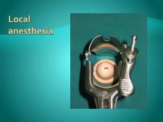
L.A Tools Dental Syringe Dental Needles
L.A Tools Local Anesthetic Cartridge
L.A Tools Local Anesthetic Cartridges Color Codes
L.A Indications • Parenteral local anesthetics are used for infiltration and nerve block anesthesia. • Because of variation in systemic absorption and toxicity, the ideal choice of local anesthetic and concentration depends on the intended procedure. • Infiltration anesthesia is often used for minor surgical and dental procedures. • Nerve block anesthesia is used for surgical, dental, and diagnostic procedures and for pain management
Nerve Block Anesthesia Mandible
Inferior Alveolar Block • Technique of choice for mandibular molars also effective for premolars, canines, and incisors. • Aim is to deposit solution around the inferior alveolar nerve as it enters the mandibular foramen
Inferior Alveolar Block Technique • The patient's mouth must be widely open. • Palpate the landmarks of external and internal oblique ridges and note the line of theptyerygomandibularraphe. • With the palpating thumb lying in the retromolarfossa, the needle should be inserted at the mid point of the tip of the thumb slightly above the occlusal plane lateral to the ptyerygomandibularraphe. • The needle is inserted ~0.5cm and if a lingual nerve block is required 0.5ml of LA is injected at this point.
Inferior Alveolar Block Technique • The syringe is then moved horizontally across the dorsum of the tongue and advanced to make contact with the lingula. • Once bony contact is made the needle is withdrawn slightly and the remainder of the LA injected. • It should never be necessary to insert the needle up to the hub. • Note that the mandibular foramen varies in position with age. • In the edentulous, the foramen, and hence the point of needle insertion, is relatively higher than in the dentate.
Additional Block (higher injection) Why? • The standard block often fails to anesthetize branches of cranial nerve V3 that originate proximal to the injection site and provide accessory innervations to the mandibular teeth. • The relatively distal location of the injection also leads to lack of anesthesia of soft tissues posterior to the mental foramen. • That why a higher injection site technique are proposed.
Gow-Gates Technique • Blocks sensation by depositing LA at head of condyle • Landmarks:– • Corner of the mouth (contralateral side) • Tragus of the ear • Disto palatal cusp of the maxillary second molar • AIMING FOR THE NECK OF THE CONDYLE
AkinosiTechnique • LA deposited above lingula • Closed-mouth technique • Does not rely on a hard-tissue landmark • Parallel to occlusal plane, height of the mucogingival junction • Advanced until hub is level with distal surface of maxillary second molar • Delayed onset of anaesthesia
Gow Gates and Akinosi • Onset is more rapid • Less effective • More accepted by patients • Pain to puncture more than Akinosi • More Effective
Mental Nerve Block The mental nerve emerges from the mental foramen lying apical to and between the first and second mandibular premolars. LA injected in this region will diffuse in through the mental foramen and provide limited analgesia of premolars and canine, and to a lesser degree incisors on that side. It will provide effective soft-tissue analgesia.
Mental Nerve Block Place the lip on tension and insert the needle parallel to the long axis of the premolars angling towards bone, and deposit the LA. Do not attempt to inject into the mental foramen as this may traumatize the nerve LA can be encouraged in by massage.
Buccal Nerve Block • The buccal nerve is not anesthetized by an inferior alveolar nerve block. • This nerve innervates the tissues and periosteum buccal to the molars, so if these soft tissues are involved in treatment, the buccal nerve should be injected as well. • The additional injection is unnecessary when treating only the teeth. • A 25 gauge long needle is recommended
Buccal Nerve Block (Continue) The needle is inserted in the mucous membrane distal and buccal to the last molar. Insert the needle to 2 to 4 mm to gently contact bone, and aspirate. If negative, slowly deposit about 1/8 of the solution in the cartridge.
Sublingual Nerve Block An anterior extension of the lingual nerve can be blocked by placing the needle just submucosallylingual to the premolars, use 0.5ml of LA.
Nerve block anesthesia Maxilla
Naso-palatine Block Anesthesia Profound anesthesia can be achieved by passing the needle through the incisive papilla and injecting a small amount of solution. This is extremely painful
Infra-Orbital Block • Rarely indicated. • A 25 gauge long needle is recommended and inserted with the bevel toward the bone in themuco-buccal fold over the first premolar. • Palpate the inferior margin of the orbit as the infra-orbital foramen lies ~1cm below the deepest point of the orbital margin. • Hold the index finger at this point while the upper lip is lifted with the thumb. • Inject in the depth of the buccalsulcus towards your finger, avoid your finger, and deposit LA around the infra-orbital nerve
Anterior Middle Superior Alveolar Block The infra-orbital nerve block does not provide adequate anesthesia to the teeth distal of the canine or if the PSA injection does not provide anesthesia for the mesiobuccal root of the first molar, an MSA block injection should be administered. A 25 gauge short needle is recommended with insertion in the muco-buccal fold by the maxillary second premolar. About ½ to 2/3 of a cartridge of anesthetic is slowly deposited at the height of the apex of the second premolar after negative aspiration
Anterior Middle Superior Alveolar Block (continue) One injection Central to second premolar, palatal and buccal soft tissue Is used to anesthetize pulp tissue and facial periodontium of the maxillary premolars and the mesio-buccal root of the first molar in some cases.
Posterior Superior Alveolar Block • The posterior superior alveolar (PSA) nerve block is a commonly used technique for achieving anesthesia for the maxillary molars. • Posterior superior alveolar block A rarely indicated technique. • The short 25 or 27 gauge needle is recommended to decrease the risk of a hematoma. • Needle is inserted distal to the upper second molar and advanced in wards, backwards, and upwards close to bone for ~2cm. • LA is deposited high above the tuberosity after aspirating to avoid the ptyerygoid plexus
Greater Palatine Nerve Block • The greater palatine nerve innervates the palatal tissues and bone distal of the canine on the side anesthetized. • Use a 27 gauge short needle with the bevel toward the palate. • Palpate the palate until the depression of the foramen is felt (usually some where medial to the second molar). • Dry the tissue, and apply antiseptic and topical anesthetic for 2 minutes. • Apply pressure with the swab for 30 seconds. Continue pressure with the swab until the injection is completed.
Greater Palatine Nerve Block (Continue) • Place the bevel against the tissue and apply pressure enough to slightly bow the needle. • Inject a few drops of anesthetic. • Release the pressure of the needle and advance the tip of the needle in to the tissues lightly. • Continue with this procedure of applying pressure to the bevel and depositing a few drops of anesthetic, then advancing, until the needle is in contact with the palatal bone. • Deposit less than a fourth to a third of a cartridge of anesthetic after negative aspiration is proven
Maxillary Nerve Block • Maxillary (V2) nerve innervates half of the maxilla, including the buccal and palatal aspects. • This injection technique issued especially in quadrant surgery or when extensive treatment is indicated for a single appointment. • It is also used when another site of injection has failed or if there is an infection in the area his technique is used more with adult patients. • It is not for the inexperienced.
MaxillaryNerveBlock(continue) • Administration through the buccal aspect involves the possibility for hematoma. • The long 25 gauge needle is recommended with the bevel of the needle facing the bone. • The needle is inserted at the mucobuccal fold near the distal of the second molar after the usual protocol of tissue preparation. • The path of the needle is similar to that of the PSA nerve block, but is inserted approximately 30mm to the pterygopalatinefossa. • Aspirate, then rotate the needle bevel ¼ turn, reaspirate. If both aspirations are negative, slowly deposit one cartridge of anesthetic (deposit ¼ then aspirate, then deposit ¼ until the entire cartridge has been administered).
Infiltrations • The aim is to deposit LA supra periosteally in as close proximity as possible to the apex of the tooth to be anaesthetized. • The LA will diffuse through periosteum and bone to bathe the nerves entering the apex. Lower concentrations of local anesthetics are typically used for infiltration anesthesia. • Variation in local anesthetic dose depends on the procedure, the degree of anesthesia required, and the individual patient's circumstances. • Reduced dosage is indicated in patients who are disabled or acutely ill, very young or very old, and in patients with liver disease, arteriosclerosis, or arterial disease.
Infiltrations Administrative techniques • The aim is to deposit LA supra periosteally in as close proximity as possible to the apex of the tooth to be anaesthetized. • Patient comfort is essential during administration of local anesthetic agents. Warming the local anesthetic solution prior to administration to 25-40° C has been recommended. • Reflect the lip or cheek to place mucosa on tension and insert the needle along the long axis of the tooth aiming towards bone
Infiltrations Administrative techniques (Continue) At approximate apex of tooth, withdraw slightly to avoid sub periosteal injection, LA Is slowly deposited. For palatal infiltrations, achieve topical analgesia first and infiltrate interdental papillae; then penetrate palatal mucosa and deposit small amount of LA under force.
Infiltration in Mandible • Buccal infiltration anesthesia in the mandible can be effective in some areas. • Indeed in children this may the preferred technique when treating the deciduous dentition. • In adult patients buccal infiltrations maybe effective in mandibular incisor region.
Local Anesthesia TechniquesPart 2 Dr. Rahaf Al-Habbab BDS. MsD. DABOMS Diplomate of the American Boards of Oral and Maxillofacial Surgery 2012