Download
1 / 17
170 likes | 173 Vues
This lecture provides an overview of Haemophilus, a group of small gram-negative rods commonly found on human mucous membranes. It discusses Haemophilus influenzae and its association with diseases, as well as the growth requirements and virulence factors of Haemophilus species. The lecture also covers other important pathogens, such as Haemophilus aegyptius and Haemophilus ducreyi.

E N D
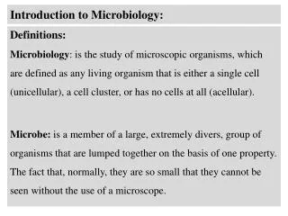
Introduction to Microbiology Anas Abu-Humaidan M.D. Ph.D. Lecture 17
Haemophilus • Haemophilae are small, sometimes pleomorphic, gram-negative rods present on the mucous membranes of humans. • Haemophilus influenzae is the species most commonly associated with disease, also Haemophilusaegyptius, and Haemophilusducreyi. • Haemophilus requires supplementation of media with one or both of the following growth-stimulating factors: (1) hemin (also called X factor for “unknown factor”) and (2) nicotinamide adenine dinucleotide (NAD; also called V factor for “vitamin”) • Haemophilus species are present in almost all individuals, primarily colonizing the mucosal membranes of the respiratory tract, H. influenzae (particularly serotype b [biotype I]) is uncommon in the upper respiratory tract. • The surface of many, but not all, strains of H. influenzae is covered with a polysaccharide capsule, and six antigenic serotypes (a through f)
Heated blood (chocolate) agar. Named for the color and contains no actual chocolate !
Haemophilus influenzae • The major virulence factor in H. influenzae type b is the antiphagocytic polysaccharide capsule, which contains ribose, ribitol, and phosphate (commonly referred to as polyribitol phosphate [PRP]). • Antibodies directed against the capsule greatly stimulate bacterial phagocytosis and complement-mediated bactericidal activity. These antibodies develop because of natural infection, vaccination with purified PRP, or the passive transfer of maternal antibodies. • When vaccines containing purified PRP antigens conjugated to protein carriers (i.e., diphtheria toxoid, tetanus toxoid, meningococcal outer membrane protein) were introduced in December 1987, a protective antibody response in infants aged 2 months and older was produced, and systemic disease in children younger than age 5 was virtually eliminated in the United States. • Most of the H. influenzae type b infections now occur in children who are not immune (because of incomplete vaccination or a poor response to the vaccine) and in elderly adults with waning immunity. • H. influenzae serotype b was responsible for more than 95% of all invasive Haemophilus infections. After introduction of the vaccine, more than half of all invasive disease is now caused by nonencapsulated (nontypeable) strains
Haemophilus influenzae • A common cause of disease in unvaccinated children (i.e., meningitis, epiglottitis [obstructive laryngitis], cellulitis). H. influenzae type b remains the most significant pediatric pathogen in many countries of the world. • Adhesins mediate colonization of the oropharynx with H. influenzae. Cell wall components of the bacteria damage the respiratory epithelium. Then translocation across both epithelial and endothelial cells occurs and the bacteria can enter the blood. • In the absence of specific opsonic antibodies directed against the polysaccharide capsule, high-grade bacteremia can develop. • Nonencapsulated strains of H. influenzae are opportunistic pathogens that can cause infections of the upper and lower airways. Most studies have shown that H. influenzae and Streptococcus pneumoniae are the two most common causes of acute and chronic otitis and sinusitis
Haemophilus influenzae • Patients with systemic H. influenzae infections require prompt antimicrobial therapy because the mortality rate in patients with untreated meningitis or epiglottitis approaches 100%. • A presumptive identification of H. influenzae can be made by the Gram stain morphology and demonstration of a requirement for both X and V factors. • The immunologic detection of H. influenzae antigen, specifically the PRP capsular antigen, is a rapid and sensitive way to diagnose H. influenzae type b disease.
Haemophilus • Haemophilusaegyptius, also called the Koch-Weeks bacillus, causes an acute purulent conjunctivitis • Haemophilusducreyican cause chancroids, a sexually transmitted disease that is most commonly diagnosed in men. Approximately 5 to 7 days after exposure, a tender papule with an erythematous base develops on the genitalia or perianal area.
Aggregatibacter • Two members of this genus are important human pathogens: A. actinomycetemcomitansand A. aphrophilus • A. actinomycetemcomitansis a Gram-negative, facultative anaerobe, non-motile bacterium that is often found in association with localized aggressive periodontitis • Both species colonize the human mouth and can spread from the mouth into the blood and then stick to a previously damaged heart valve or artificial valve, leading to the development of endocarditis.
Pasteurella • Pasteurella is a genus of Gram-negative, facultatively anaerobic, fermentative coccobacilli commonly found as commensals in the oropharynx of healthy animals. • Most human infections result from animal contact (e.g., animal bites, scratches, shared food). • Pasteurella multocida(the most common isolate) and Pasteurella canisare human pathogens.
Vibrio • Gram-negative, facultatively anaerobic, fermentative rods, characterized by apositive oxidase reaction and the presence of polar flagella . • Vibrio species can grow on a variety of simple media within a broad temperature range (from 14° C to 40° C). And tolerate a wide range of pH (e.g., pH of 6.5 to 9.0) but are susceptible to stomach acids. • All species of Vibrio require sodium chloride (NaCl) for growth. Most species are halophilic (“salt-loving”). • Vibrio species, including V. cholerae, grow naturally in estuarine and marine environments worldwide. • Pathogenic vibrios can also flourish in waters with chitinous shellfish
Vibrio • All strains possess lipopolysaccharides consisting of lipid A (endotoxin), core polysaccharide, and an O polysaccharide side chain. • The O polysaccharide is used to subdivide Vibrio species into serogroups, V. cholerae O1 and O139 produce cholera toxin and are associated with epidemics of cholera. Other strains of V. cholerae generally do not produce cholera toxin and do not cause epidemic disease. • Cholera is spread by contaminated water and food rather than direct person-to-person spread, because a high inoculum (e.g., >108 organisms) is required to establish infection in a person with normal gastric acidity. • Cholera is usually seen in communities with poor sanitation. Immunoassays for the detection of cholera toxin or the O1 and O139 lipopolysaccharides are used for the diagnosis of cholera in endemic areas. • It is estimated that 3 to 5 million cases of cholera and 120,000 deaths occur worldwide each year. Seven major pandemics of cholera have occurred since 1817, resulting in thousands of deaths and major socioeconomic changes.
Vibrio • The majority of individuals exposed to toxigenic V. cholerae O1 have asymptomatic infections or self-limited diarrhea; however, some individuals develop severe, rapidly fatal diarrhea. V. cholerae O1 does not produce a capsule, so infections with this organism do not spread beyond the confines of the intestine. • The clinical manifestations of cholera begin an average of 2 to 3 days after ingestion of the bacteria (can be <12 hours), with the abrupt onset of watery diarrhea and vomiting. Fever is rare. • The feces-streaked stool specimens become colorless and odorless, free of protein, and speckled with mucus (“rice-water” stools). • Patients with cholera must be promptly treated with fluid and electrolyte replacement before the resultant massive fluid loss leads to hypovolemic shock.
Vibrio cholera • Virulence of V. cholerae involved acquisition of first a sequence of genes including the toxin co-regulated pilus (TCP) on what is termed the vibrio pathogenicity island (VPI-1), followed by infection with the bacteriophage CTXΦthat encodes the genes for the two subunits of cholera toxin (ctxAand ctxB). • TCP serves as the cell surface receptor for the bacteriophage, permitting it to move into the bacterial cell, where it becomes integrated into the V. cholerae genome.
Vibrio cholera • The cholera toxin is a complex A-B toxin. The active portion of the A subunit is internalized and interacts with G proteins that control adenylate cyclase, leading to the catabolic conversion of adenosine triphosphate (ATP) to cyclic adenosine monophosphate (cAMP). This results in a hypersecretion of water and electrolytes. • The resulting severe fluid and electrolyte loss can lead to dehydration, painful muscle cramps, metabolic acidosis (bicarbonate loss), and hypokalemia and hypovolemic shock (potassium loss), with cardiac arrhythmia and renal failure. • The mortality rate is as high as 70% in untreated patients but less than 1% in patients who are promptly treated with replacement of lost fluids and electrolytes
Further reading: • Murray - Medical Microbiology 8th Edition • Section 4: Bacteriology • Chapter 24: • Chapter 26: