Download
1 / 36
381 likes | 649 Vues
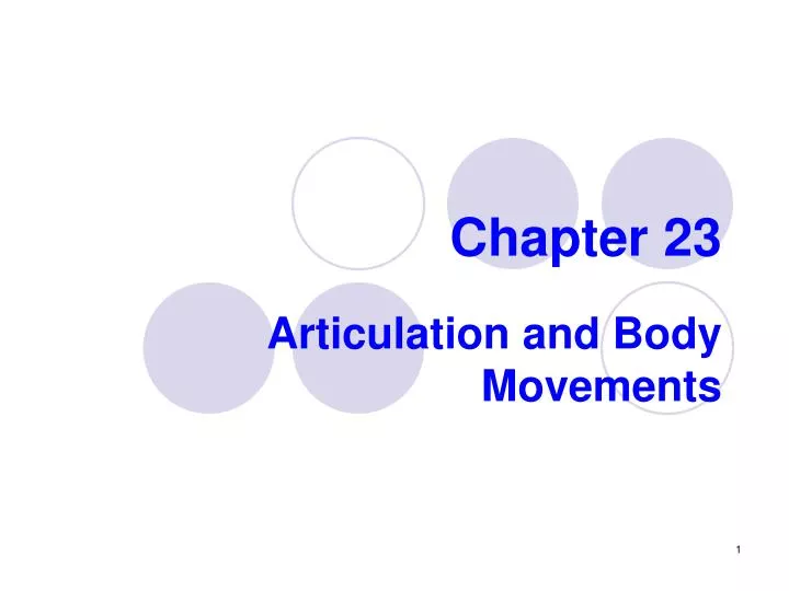
Chapter 23. Articulation and Body Movements. Functions of articulations. Articulations Where two bones interconnect To hold bones together To allow movements of the body. Functional classification. Immovable joints- Synarthroses More predominant in the axial skeleton

E N D
Chapter 23 Articulation and Body Movements
Functions of articulations • Articulations • Where two bones interconnect • To hold bones together • To allow movements of the body
Functional classification • Immovable joints- Synarthroses • More predominant in the axial skeleton • Slightly moveable joints- Amphiarthroses • More predominant in the axial skeleton • Freely moveable joints- Diarthroses • More predominant in the appendicular skeleton
Structural classification • Fibrous joints • No presence of joint cavity • They are synarthroses or amphiarthroses • Fibrous tissue present • Suture = skull bones bound together by dense connective tissue. It is a synarthrose. Bones interlock • Gomphosis= teeth bound to bony sockets by periodontal ligaments
Structural classification • Synostosis = two bones completely fused. Portions of the skull • Syndesmosis = bones connected by a ligament. Distal articulation between fibula and tibia. Movement varies from immovable to slightly variable.
Fibrous Structural Joints: Syndesmoses Figure 8.1b
Cartilaginous joints • Bones connected by a pad or plate of cartilage • Symphysis = bone separated by fibrocartilage. Pubic symphysis and intervertebral joints. It is amphiarthrotic • Syncondrosis= bones connected by hyaline cartilage. Epiphyseal plate and articulation of the first rib with the sternum. It is synarthrotic
Synovial joints • Bony surfaces enclosed within articularcapsule(dense connective tissue) • Synovial membrane-inside of the capsule • Secretes the synovial fluid • Synovial cavity • Articular cartilage • Resemble hyaline cartilage and covers the bone ends
Synovial joints • Meniscior articular discs • Improves the fit of the joint • Minimizes the wear and tear of the joint • Fat pads • Bursae and tendon sheath • Synovial sacs between tendons • They reduce friction • May or may not be present in the joint
Synovial joints • Reinforcing ligaments • Intrinsic or capsular- it is a thickening part of the caspsule • Extracapsular- outside of the capsule • Intracapsular- inside of the capsule
Synovial Joints: Stability • Stability is determined by: • Articular surfaces – shape determines what movements are possible • Ligaments – unite bones and prevent excessive or undesirable motion • Muscle tone
Structural Classification of the Synovial Joints • Plane - articular surface is flat or slightly curved • Hinge– round process of one bone fits into the concave surface of the other bone. Elbow • Pivot- allows rotational movement between two bones. • Condyloid – convex surface articulating with a concave one
Structural Classification of the Synovial Joints • Saddle -one concave and one convex bone facing it other • Ball-and-socket - permit rotation and other movements
Types of movements of synovial joints • Gliding • Flexion • Extension, hyperextension • Abduction • Adduction • Rotation • Circunduction • Elevation • Depression
Types of movements of synovial joints • Pronation • Supination • Inversion • Eversion • Dorsiflexion • Plantar flexion • Protraction • Retraction • Opposition
Selected synovial joints- Knee • Menisci • Act as cushion • Provide lateral stability to the joint • Lateral and medial • Bursae
Knee joint • Collateral ligaments • Prevent rotation during extension • Reinforce the sides of the knee • Medial or tibial • Lateral or fibular
Knee joint • Cruciate ligaments • Prevent anterior-posterior displacement of the joint, overflexion and hyperextension of the joint • Anterior • Posterior
Knee joint • Popliteal ligaments • Reinforce the posterior surface of the knee • Patellar ligament-from patella to the tibia • Patellar retinaculum • Lateral and medial • Merge with the capsule • Patellar and retinaculum ligaments support the anterior surface of the knee
The Knee Joint Figure 9.12c, d
Hip joint • Ball and socket diarthroses • Acetabular labrum • Circular rim of fibrocartilage. Deepens the socket
Hip joint • Ligamentumteres or ligament of the head of the femur • From fovea capitis to the acetabulum. • Helps to secure the femur • Iliofemoral ligament • Pubofemoral ligament • Ischiofemoral ligament
Temporomandibular joint (TMJ) • Between mandibular fossa and mandibular condyle • Articular disc • Divides the joint in superior and inferior compartment • Lateral ligament
Joint Disorders • Sprain • Damage of the ligament by excessive stretch or tear. Slow and painful healing • Dislocation • Bones are forced out of their normal position • Reduction
Joint Disorders • Adhesion • Fibrous bands between the surfaces where the bones meet • Spurs • Extra bone growing along the joint • Bursites • Damage or inflamation of the bursa by blow or friction
Osteoarthritis (OA) • Most common chronic arthritis; often called “wear-and-tear” arthritis • Affects women more than men • More prevalent in the aged, and is probably related to the normal aging process
Arthritis • Gouty Arthritis • Rheumatoid Arthritis