Download
1 / 28
431 likes | 1.12k Vues
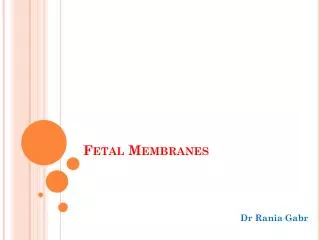
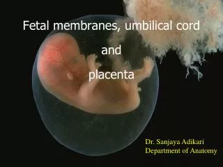
Fetal Membranes. Dr. Mohammad Saeed Vohra. vohra@ksu.edu.sa. OBJECTIVES. By the end of the lecture the student should be able to: List the components of the fetal membranes. Describe the stages of development of the components. Describe the structure and function of the components.

E N D
Fetal Membranes Dr. Mohammad Saeed Vohra • vohra@ksu.edu.sa
OBJECTIVES • By the end of the lecture the student should be able to: • List the components of the fetal membranes. • Describe the stages of development of the components. • Describe the structure and function of the components. • Describe their fate and the possible congenital anomalies.
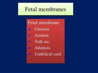
Fetal Membranes Contents Functions Umbilical cord (Connecting Stalk) Amnion Yolk sac Allantois Protection Nutrition Respiration Excretion Hormone
Components of Fetal Membrane • Umbilical cord (Connecting Stalk) • Amnion • Amniotic Fluid • Yolk Sac • Allantois
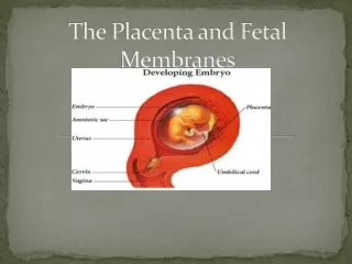
Umbilical Cord • It is a pathway which connects the ventral aspect of the embryo with the chorion or placenta • It is a soft tortuous cord which measures from 30- 90 cm long (average 55) and about 1-2 cm. in diameter • It has a smooth surface because it is covered by epithelial layer of the amnion
Structure of Umbilical Cord • It is composed of: • 1-Connecting stalk: • Itcontains Alantois + 4 Umbilical vessels (2-arteries + 2-veins),after folding of embryo, it lies on ventral aspect of embryo • Its extra embryonic mesoderm becomes modified to form Wharton’s jelly • 2-Yolk stalk (Vitello-intestinal duct): • A narrow, elongated duct which connects gut to yolk sac • It contains Vitelline Vessels • As development proceeds, it is obliterated and the vitelline vessels disappear
Attachment of Umbilical Cord • Normally, it is attached to a point near the centre of the fetal surface of the placenta
Anomalies of Umbilical Cord • (1) Abnormal Attachment of cord • a-Battledore placenta : • May take the marginal attachment to placenta (it is not dangerous). • b-Velamentous insertion of the cord : • UC is attached to amnion away from placenta, (It is dangerous to foetus due to rupture of blood vessels during labour)
Anomalies of Umbilical Cord • (2) Abnormalities in Length: • a-Very Long Cord: • it is dangerous , it may surround neck of fetus and cause its death. • b-Very Short Cord: • it is dangerous because it may cause premature separation of placenta, or the cord itself may rupture
Anomalies of Umbilical Cord • (3) False and True knots of umbilical cord:a-False knots: • UC looks tortuous due to twisting of umbilical vessels (umbilical vessels are longer than the cord), these knots are normal and do not cause any harm to fetus • b-True knots: • Are rare (1%) of pregnancy, but very dangerous because they may cause obstruction to blood flow in umbilical vessels, leading to fetal anoxia & fetal death True Knots in 20-weeks fetus
Amnion • It is a thin, transparent & tough fluid-filled, membranous sac surrounding the embryo.
Amnion • At First : It is seen as a small cavity lying dorsal to embryonic plate. • At Stage of Chorionic Vesicle: The amnion becomes separated from the chorion by chorionic cavity or E.E.coelom. • After Folding: the amnion expands greatly and is now located on the ventral surface of embryo. • As a result of expansion of the amnion, extra embryonic coelom is gradually obliterated and amnion forms the epithelial covering of umbilical cord.
Amniotic Fluid • It is a watery fluid inside the amniotic cavity, that plays a major role in fetal growth & development • It increases slowly, reaching about 700-1000 ml by full term of 37 weeks.
Sources of amniotic fluid • It has Fetal & Maternal Sources: • Initialy, some amniotic fluid is secreated by amniotic cells. • Most of fluid is derived from Maternal tissue by: • 1-Diffusion across amnio-chorionic membrane from placenta. • 2-Diffusion across chorionic plate (chorionic wall related to placenta) from the maternalblood in intervillous spaces. • Later, it is derived from Fetus through: • Skin, Fetal Respiratory tract & mostly by excreting Urine (at beginning of 11th week)
Functions of amniotic fluid • Provides symmetrical external growth of embryo • Acts as a barrier to infection (it is an aseptic medium) • Permits normal fetal lung development • Prevents adherence of embryo to amnion • It protects embryo against external injuries • Keeps the fetal body temperature constant • Allows the embryo to move freely, aiding muscular development in the limbs • It is involved in maintaining homeostasis of fluid & electrolytes
Composition of amniotic fluid • 99% of amniotic fluid is water • It contains un-dissolved material of desquamated fetal epithelial cells + organic + inorganic salts • As pregnancy advances, composition of amniotic fluid changes as fetal excreta (meconium = fetal feces/& urine) are added • Amniotic fluid removed by amniocentesis permits studies on fetal enzymes, hormones and diagnosis of fetal sex and chromosomal abnormalities
Circulation & Fate of amniotic fluid • Amniotic fluid remains constant & in balance • --Most fluid is swallowed and few passes into lungs by fetus, and absorbed into fetal blood, whereit is metabolised • --Part of fluid passes through placental membrane into maternal blood in intervillus space, • Other part of fluid is excreted by fetal kidneys into amniotic sac
Anomalies of Volume of Amniotic Fluid • (1) Oligohydramnios: • The volume is less than ½ litres • Causes of oligohydramnios: • Placental insufficiencywith low placental blood flow • Preterm rupture of amnio-chorionic membrane occurs in 10% of pregnancies • Renal Agenesis(failure of kidney development), or Obstructive Uropathy(urinary tract obstruction) lead to absence of fetal urine (the main source) • Complications of oligohydramnios: • Fetal abnormalities (pulmonary, facial & limb defects)
Anomalies of Volume of Amniotic Fluid • (2) Polyhydramnios (Hydramnios): • The volume is more than 2 litres,Ultrasonography is the choice technique for diagnosis of hydramnios. • Causes • Fetal ( 1-20% ) : Esophageal atresia. • Maternal (2-20%) : defects in maternal circulations. • Idiopathic (3-60%)
Yolk Sac • Although it is called ‘Yolk’ sac, it does not contain any yolk, but it is essential in the transfer of nutrients to embryo during 2nd & 3rd weeks, when the uteroplacental circulation is not established
Stages of development • Primary Yolk Sac • Appears in Blastocyst stage at 10-days, it lies ventral to embryonic plate. Its roof is formed by hypoblast (primary endoderm), its wall is formed by exocoelomic membrane, which lines inner surface of cytotrophoblast, then retracts from the trophoblast due to formation of extraembryonic mesoderm
Stages of development • Secondary Yolk Sac: • Appears in chorionic vesicle stage • Its roof is formed by hypoblast (embryonic endoderm), its wall is formed by exocoelomic membrane + inner layer (splanchnic) of extraembryonic mesoderm. • At day 16, it sends a diverticulum from its dorsocaudal end into substance of connecting stalk (Allantois)
Definitive Yolk Sac • After folding, part of Yolk Sac is enclosed in the embryo to form the gut (foregut, midgut & hindgut). • The remainder of Yolk Sac outside the embryo becomes the Definitive Yolk Sac • The midgut is temporarily connected to Definitive Yolk Sac by a narrow duct: Vitello-intestinal duct(Yolk stalk),which is incorporated inside umbilical cord. then obliterated, fibrosed and degenerated by end of (6th week).
Functions of Yolk Sac • Early in 3rd week,Blood developmentfirst occurs in the extra-embryonic mesoderm which covers wall of yolk sac, until hemopoietic activity begins in the liver during 6thweek • During 4th week, endoderm of yolk sacis incorporated into embryo to form primordial gut • It gives rise also to epithelium of Respiratory & G.I.T. In 3RD week: Primordial germ cells originate in endodermal lining of the wall of caudal end of yolk sac migrate into developing sex glands to differentiate into germ cells (spermatogonia or oogonia)
Fate of Yolk Sac • Yolk stalkdetached from midgutby the end of 6th week. In about 2% of adults, its proximal intra-abdominal part persists as ilealdiverticulum(Meckeldiverticulum). • At 10 weeks,small definitive yolk sac lies in chorionic cavity between amniotic & chorionic sacs • At 20 weeks, as pregnancy advances, definitive yolk sacatrophies and becomes very small cyst. • In unusual cases, it persists under amnion near the attachment of Umbilical cord, on fetal surface of placenta. Its persistence is of no significance
Allantois • In 3rd week, it appears as a diverticulumfrom caudal wall of yolk sac that extends into connecting stalk. • During 2nd month, the extra- embryonic part of allantoisdegenerates • During 3rd month, Intra-embryonic part of allantois extends from urinary bladder to umbilical cord as thick tube , ‘urachus’ • After birth, the urachus is obliterated and fibrosed to form median umbilical ligament, that extends from apex of urinary bladder to umbilicus.
Functions of Allantois • Blood formation in its wall during 3rd to 5th week. • Its blood vessels persist as umbilical vein & arteries.