Download
1 / 8
560 likes | 2.28k Vues
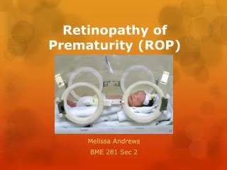
Retinopathy of prematurity (ROP). Proliferative retinopathy Affects pre -term infants exposed to high ambient oxygen concentrations

E N D
Retinopathyofprematurity(ROP) • Proliferativeretinopathy • Affectspre-term infantsexposed to high ambient oxygen concentrations • After 8 monthsofgestationretinalvesselsreachthenasalperipheryof retina, althoughthey do not reachthetemporalperipheryuntil 1 monthafterdelivery • Incompletelyvascularizedtemporal retina issusceptible to oxygen damage
Clinical features Theseverityof ROP canbedeterminedaccording to location, extent, stages and „plus“ disease. Locationisdeterminedaccording to 3 zonescentred on theopticdisc. • Zone 1isbounded by theimaginarycirclewhosradiusistwicethe distance fromthedisc to themacula • Zone 2extendsfromtheedgeofzone 1 to a point tangential to thenasaloraserrata and round to an area neartemporalequator • Zone 3consistsof a residualtemporalcrescentanterior to zone 2
Clinicalfeatures5 stages Stagingis a follows: • Stage 1 = demarcation line. Thin, tortuous, grey-white line whichrunsparallelwiththeoraserrata. The line separatestheavascularimmatureperipheral retina fromthevascularposterior retina. • Stage 2 = ridge. The demarcation line develops into a ridge of tissue, which extends out of the plane of the retina. The ridge represents a mesenchymal shunt which joins veins with arteries.
5 stages • Stage 3 = ridge with extraretinal fibrovascular proliferation. Retinal and vitreous haemorrhage also develop. • Stage 4 = subtotal retinal detachment. Progression of fibrovascular proliferation give rise to a tractional detachment. • Stage 5 = total retinal detachment.
„Plus“ disease Is characterized by dilatation of the veins and tortuosity of the arterioles in the posterior fundus. When these changes are present, a „plus“ sign is added to the stage number.
Screening • Examination of the retina in all infants born at less than 36 months or weighing less than 1500g, who have received supplemental oxygen • The pupils in a pre-term infant should be dilated (2,5% phenylephrine)
Treatment • Ablation of avascular immature retina by either cryotherapy or laser photocoagulation (stage 3) • Scleral buckling with or without PPV (stage 4,5)