Download
1 / 1
10 likes | 118 Vues
Abstract # 2666. BT474 MCF10A - + - + BN108. PARP-1. Timosaponin A3 is a steroidal saponin from Anemarrhena asphodeloides that has a selective cytotoxic activity towards cancer cells

E N D
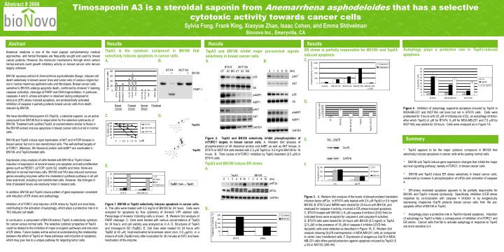
Abstract # 2666 BT474 MCF10A - + - +BN108 PARP-1 Timosaponin A3 is a steroidal saponin from Anemarrhena asphodeloides that has a selective cytotoxic activity towards cancer cells Sylvia Fong, Frank King, Xiaoyue Zhao, Isaac Cohen, and Emma Shtivelman Bionovo Inc., Emeryville, CA Results Results Results Results Abstract Results Autophagy plays a protective role in TspA3-induced apoptosis Figure 4 .Inhibition of autophagy augments apoptosis induced by TspA3 in MDA-MB-231 and MCF10A cell lines but not in BT474 cells. Cells were pretreated for 3 hours with 20 M of chloroquine (CQ), an autophagy inhibitor, after which TspA3 (4 M for BT474, 5 M for MDA-MB-231 and 7.5 M for MCF10A) was added for 24 hours. Cells were analyzed as in Figure 1A. ER stress is partially responsible for BN108- and TspA3-induced apoptosis C. D. E. F. Figure 3 .A. Western blot analysis of the levels of phosphorylated translation initiation factor eIF2α, in BT474 cells treated with 2.5 M TspA3 or 0.5 mg/ml BN108. B. BT474 and IMR90 were treated for 24 hours with BN108, and analyzed for caspase 4 activity, involved in ER stress-induced apoptosis. C. BT474 treated with BN108 (± 8 M caspase 4 inhibitor LEVD-fmk) for indicated times were analyzed for caspase-4 and caspase-9 activities. D. BT474 cells were treated for 24 hours with BN108 (0.5 mg/ml) or TspA3 (2.5 M) in the presence of 10 M of caspase 4 inhibitor LEVD-fmk. Apoptotic cells were detected as described in Figure 1A.. E. Western blot analysis showing Grp78 overexpression in MDA-MB-231 cells as compared to vector (vec) transfected cells. F. Expression of exogenous Grp78 in MDA-MB-231 cells offers partial protection against apoptosis induced by TspA3 (5 M) or MG132 (250 nM). Botanical medicine is one of the most popular complementary medical approaches, and herbal therapies are frequently sought and used by breast cancer patients. However, the molecular mechanisms through which certain herbal extracts exert growth inhibitory activity on breast cancer cells remain largely unknown. BN108, aqueous extract of Anemarrhena asphodeloides Bunge, induces cell death selectively in breast cancer lines and tumor cells of various origins but not in normal mammary epithelial cells and fibroblasts. Breast cancer cells sensitive to BN108 undergo apoptotic death, confirmed by Annexin V staining, caspase activation, cleavage of PARP and DNA fragmentation. In particular, caspases 4 and 9, whose activation is observed during endoplasmic reticulum (ER) stress induced apoptosis, are proteolytically activated. Inhibition of caspase 4 partially protects breast cancer cells from death induced by BN108. We have identified timosaponin A3 (TspA3), a steroidal saponin, as an active compound from BN108 that is responsible for the selective cytotoxicity of BN108. Treatment with purified TspA3 at concentrations similar to those in the BN108 extract induces apoptosis in breast cancer cells but not in normal cells. BN108 and TspA3 induce rapid inactivation of AKT and mTOR kinases in breast cancer but not in non-transformed cells. The well-defined targets of mTORC1, S6kinase, S6 ribosomal protein and 4eBP1 are inactivated in BN108- and TspA3-treated cells. Expression array analysis of cells treated with BN108 or TspA3 shows induction of expression of several known pro-apoptotic and anti-proliferative genes such as REDD1, p21CIP, cyclin G2, stratifin and more. None are affected in normal mammary cells. BN108 and TA3 also induced numerous genes encoding enzymes within the cholesterol synthesis pathway in all cell lines examined, including non-transformed cells. However, the changes in total cholesterol levels are relatively minor in treated cells. In addition, BN108 and TspA3 induce pattern of gene expression consistent with induction of ER stress and authophagy. Inhibition of mTORC1 and induction of ER stress by TspA3 are most likely contributing to the activation of autophagy, which plays a protective role in in TA3 induced cell death. In conclusion, a component of BN108 extract, TspA3 is selectively cytotoxic for cancer versus normal cells. The selective cytotoxic properties of TspA3 could be related to the inhibition of major oncogenic pathways and induction of ER stress. Future studies will be aimed at understanding the relationship between the effect of TspA3 on these pathways and induction of apoptosis, which may give rise to a unique pathway for targeting tumor cells. • TspA3 is thecytotoxic compound in BN108 that selectively induces apoptosis in cancer cells. • A.B. • D. • E. • Figure 1. BN108 or TspA3 selectively induces apoptosis in cancer cells. A. The cells were treated with 0.5 mg/ml of BN108 for 24 hours. Cells were analyzed for apoptosis by flow cytometry of Annexin V/PI stained cells. Percentage of Annexin V-binding cells is shown. B. Western blot analysis of PARP cleavage. C. Cells were treated with various concentrations of TspA3 for 24 hours, and cell viability was analyzed as in A. D. Structures of TspA3 and timosaponin B2 (TspB2). E. Cell lines were treated for 24 hours with TspB2 at 40 M, heat-inactivated laminarinase alone (lam, 0.5 g/ml), or a mixture of both (TspB2+lam) after incubation for 30 minutes at 500C and heat-inactivation of the enzyme. TspA3 and BN108 inhibit major pro-survival signals selectively in breast cancer cells. A. B. . BT474 MCF10A UT A3 BN UT A3 BN 0 1 4 8 16 (Hrs) pAKT AKT pS6 S6 p4eBP1 4eBP1 GAPDH pS6 S6 p4eBP1 4eBP1 pAKT AKT GAPDH BreastProstateNormalFibroblast Cancer CancerBreast TspA3 Summary Figure 2. TspA3 and BN108 selectively inhibit phosphorylation of mTORC1 targets in breast cancer cells. A. Western blot analysis of phosphorylation of s6 ribosomal protein and 4eBP, as well as AKT kinase, in BT474 or MCF10A cells treated with 2.5 M TspA3 or 0.5 mg/ml BN108 for 16 hours. B. Time course of mTORC1 inhibition by TspA3 treatment (2.5 M) in BT474 cells. Vec Grp78 (mM) Grp78 TspB2 • TspA3 appears to be the major cytotoxic compound in BN108 that selectively induces apoptosis in cancer cells while sparing normal cells. • BN108 and TspA3 induce gene expression changes that inhibit the major survival signaling pathway, namely mTORC1, in breast cancer cells. • BN108 and TspA3 induce ER stress selectively in breast cancer cells, evidenced by increase in phosphorylation of eIF2α and activation of caspase 4. • ER-stress mediated apoptosis appears to be partially responsible for BN108- and TspA3- induced cytotoxicity. Specifically, inhibition of ER stress response by co-incubation with caspase 4 inhibitor or by exogenously expressing chaperone Grp78 protects breast cancer cells from the pro-apoptotic effect of BN108 or TspA3. • Autophagy plays a protective role in TspA3-induced apoptosis. Induction of autophagy by TspA3 is likely a consequence of inhibition of mTORC1 and ER stress. Cancer cells that fail to activate autophagy in response to TspA3 are more sensitive to it. Vec Grp78 GAPDH TspA3 and BN108 induce ER stress A. B. TspA3 BN108 0 1 2 4 8 16 1 2 4 8 16 (hrs) TspB2 + lam→ TspA3 peIF2α eIF2α GAPDH References