Download
1 / 48
510 likes | 797 Vues
2013 PARASITOLOGY WORKSHOP . LYNNE S. GARCIA, MS, FAAM, CLS, BLM Diagnostic Medical Parasitology Workshop 2013 UPDATE – PART 5 OTHER BLOOD PARASITES SPONSORED BY MEDICAL CHEMICAL CORPORATION www.med-chem.com. Babesia spp. Ixodes scapularis. BABESIA SPP. Babesia divergens : Europe

E N D
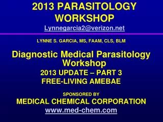
2013 PARASITOLOGY WORKSHOP LYNNE S. GARCIA, MS, FAAM, CLS, BLM Diagnostic Medical Parasitology Workshop 2013 UPDATE – PART 5 OTHER BLOOD PARASITES SPONSORED BY MEDICAL CHEMICAL CORPORATION www.med-chem.com
Ixodes scapularis BABESIA SPP. • Babesia divergens: Europe • Rare, but 42% mortality • Babesia microti: US, parts of Europe, Japan • Northeast, south to New Jersey; 5% mortality • Most cases mild; more severe in immunosuppressed • WA1, CA1, MO1 (Babesia divergens-like), states • Very serious in splenectomized patients • Related to canine pathogen, B. gibsoni • WA1, CA5: Now Babesia duncani • More pathogenic than B. microti
Babesia spp.Clinical Disease • Incubation period: 1-3 weeks • Symptoms: sudden onset (B. divergens) with shock; fever, chills, confusion, jaundice • Symptoms: Mild and resolve on their own (B. microti); severe cases fulminating malaria-like symptoms (malaise, chills, myalgia, anemia, fatigue, fever, hepato- and splenomegaly); hemolytic anemia, particularly in asplenic individuals (days to months) • Severe complications: multiorgan dysfunction, renal failure, coma, death
LABORATORY DIAGNOSISKEY POINTS - BABESIA • Draw blood, prepare blood films immediately • Examine 200-300 oil immersion fields on BOTH thick and thin films • Wright’s, Wright-Giemsa, Giemsa, or a rapid stain can be used (original morphology described from Giemsa stain); if WBCs look OK, then any parasites will look OK; color variation is normal • Parasites may be missed using automation • One set of negative blood films does NOT rule out Babesia; if positive, quantitate organisms present
Other blood parasites: Babesia spp. ArtifactsPlasmodium falciparum
LEISHMANIASIS – PARASITIC FORMS • Amastigote: round or oval, about 2-5µm, intracellular, prominent nucleus and dark staining bar-shaped structure called • Kinetoplast: includes very short flagellum • Promastigote: in the insect vector, elongate with flagellum projecting from anterior end
LEISHMANIASIS – KEY FACTS~350 Million in 88 countries at risk • Cutaneous: macrophages of skin • Old World: Leishmania tropica, L. major, L. aethiopica • New World: L. mexicana, L. braziliensis • Mucocutaneous:macrophages of skin and buccal cavity or nasopharynx • Leishmania braziliensis • Visceral:macrophages of spleen, liver, bone marrow (reticuloendothelial system) • Leishmania donovani
CUTANEOUS LEISHMANIASIS • Transmitted via bite of sandfly (Phlebotomus, Lutzomyia) • Multiplication of amastigotes may last months to life; usually confined to skin • Skin lesions = raised papules, ulcers on exposed parts of body • Parasite receptors to collagen may explain tropism for skin –abundant component of matrix 10
CUTANEOUS LEISHMANIASIS IN U.S. MILITARY PERSONNEL 2002 - 2008 • Afghanistan, Iraq, Kuwait: >1000 cases • Cases isoenzyme analysis: L. major • Consider cutaneous leishmaniasis withskin lesions (Southwest/Central Asia) • Almost all with CL infected in Iraq • Decrease risks • Improve living conditions, heighten awareness, protective measures (permethrin- treated clothes, bed nets; exposed skin, repellent 30-35% DEET, dusk to dawn, vector control)
Leishmaniasis Control Program • Program begun in 2003 (high numbers of sand flies, soldiers receiving many bites per night, 1.5% of female flies infected) • Risk assessment overall • Enhancement of use of personal protective gear • Vector and reservoir control • Education of military personnel about sand flies and leishmaniasis Coleman, RE, 2006. J Med Entomol 43:647-62.
CUTANEOUS LEISHMANIASIS : CLINICAL PRESENTATION • First set of patients: median age = 29 • Deployment ~ 60 days before noting skin lesions (range of 21 to 150 days) • Median of 3 (1 to 9) skin lesions (upper and lower extremities); painless, enlarged slowly, central ulceration, often covered with eschar, surrounded by erythematous, indurated border • No patients had systemic symptoms • Microscopy of tissue, culture, isoenzyme analysis • Leishmania major
CUTANEOUS LEISHMANIASIS: DIAGNOSIS • Organism isolation by smear or culture; histology • Lesion sampling: dental broach, slit scrape method, aspiration of lesion edge (multiple samples required), biopsy • Tissue sampling better than other smearing techniques; culture positive in 80% of cases • LD bodies seen in about 30% of patients; many lymphocytes/plasma cells in wet ulcerative lesions; dry lesions = granulomas / fewer cells
Culture LEISHMANIASIS Specimen Collection
CUTANEOUS LEISHMANIASIS : THERAPY • Since 1978: Walter Reed Army Medical Center • Pentavalent antimony: sodium stibogluconate (Pentostam, Wellcome Foundation) • Not licensed in U.S., provided by WRAMC • Treatment: 20 mg/kg body weight/day; side effects, fatigue, arthralgia, myalgia, headache, chemical pancreatitis • Microscopy of tissue, culture, isoenzyme analysis • Leishmania major
MUCOCUTANEOUS LEISHMANIASIS • Transmitted via bite of sandfly (Phlebotomus, Lutzomyia) – Leishmania braziliensis • Multiplication of amastigotes: months to life; 18 mo for self-cure • Nasal, pharyngeal, buccal mucosal lesions = years after initial cutaneous lesion • ML begins with erythema, edema, inflammation, stuffiness, difficulty breathing, occasional bleeding: loss of lips, parts of nose and palate
MUCOCUTANEOUS LEISHMANIASISL. mexicana Complex • Common throughout Mexico, Belize, Guatemala and southern United States (Arizona, Texas) • Forest rodents (wood rats) are important hosts • Prolonged exposure = “chicleros” live in forest collecting chewing gum latex (30% infected in first year), timber cutters, road builders, farm workers • Two culture-positive and four PCR-pos rodents Leishmania-positive. Isolates extend geographic and ecologic range of enzootic leishmaniasis in the United States and represent a new host record. Now in Tucson, Arizona.
VISCERAL LEISHMANIASIS(Kala Azar) • Amastigotes in macrophages disseminate to liver, spleen, and bone marrow; hepatomegaly and splenomegaly • Protective Th-1 responses are reduced or absent • Hypergammaglobulinemia present but does not clear the infection
VISCERAL LEISHMANIASIS(Kala Azar) • Transmitted via bite of sandfly (Phlebotomus, Lutzomyia) – Leishmania donovani • Patients who have recovered may develop skin lesions – post kala-azar dermal leishmaniasis • Cell mediated responses absent = develop after successful treatment • VL begins with fever (double peaks) –hepatosplenomegaly; weight loss, nose bleeds, diarrhea, cough; elevated gammaglobulins
LEISHMANIASISCure – Yes or No? • Currently, no reliable way to assess cure • Most researchers think sterile cure is unlikely and/or uncommon • Evidence: • Reactivation years later (immunosuppressed or “traumatized” persons & animals) • Demonstration of organisms years after exposure, self-healing, or treatment • Persistence of immunologic markers • Extrapolation of information about other intracellular pathogens
OTHER BLOOD PARASITES(Trypanosomes) • African Trypanosomiasis, T. brucei gambiense* (West), T. brucei rhodesiense (East) Tsetse fly • African Sleeping Sickness (chronic*, CNS disease) • Acute or chronic disease, blood, buffy coat, CSF • American Trypanosomiasis, Trypanosoma cruzi • Triatomid bug (feces), blood, shared needles, congenital, organ transplants • Myocarditis, GI tract, acute or chronic disease • Blood, buffy coat, culture, xenodiagnosis • Trypanosoma rangeli (like T. cruzi) • Nonpathogen, but overlaps geographic range
TRYPANOSOMIASIS, AFRICAN • Caused by Trypanosoma brucei gambiense (West) and T. b. rhodesiense (East) • Transmitted by bite of tsetse fly (Glossina spp.) • T. b. gambiense in West, Central Africa by rivers and streams; animal reservoirs insignificant • T. b. rhodesiense in East, Central Africa in the savanna; wild game major reservoir hosts • After initial development in skin, trypomastigotes found in blood, lymph notes, and CSF
TRYPANOSOMIASIS, AFRICAN – CLINICAL DISEASE • Primary lesion may develop at bite site - chancre • Fever earliest sign of generalized infection • T. b. gambiense - after initial development in skin, trypomastigotes in blood, – fever delayed months to years; chronic – onset of CNS slow – death often > year after infection • T. b. rhodesiense – fever begins days to weeks; rapidly progressive illness – may die of myocarditis before CNS is invaded; patients often die of infection, including pneumonia
TRYPANOSOMIASIS, AFRICAN – LABORATORY DIAGNOSIS • Trypomastigotes in early cases with no evidence of CNS disease • If lymphadenopathy, lymph node aspiration; if CNS involved, centrifuged CSF; detection of IgM in CSF; very high gamma globulins – variant surface glycoproteins (VSG); antigenic variation • T. b. gambiense – may be more difficult to diagnose; fewer trypomastigotes seen • T. b. rhodesiense – higher parasitemia seen
SUMMARY • T. b. gambiense and T. b. rhodesiensetrypomastigotes are found in the blood • West African disease more chronic – progresses to actual sleeping sickness with invasion of the CNS • East African disease more acute – often leads to death within a year prior to typical CNS invasion • T. b. gambiense and T. b. rhodesiense (animal reservoirs) transmitted by the tsetse fly.
AMERICAN TRYPANOSOMIASIS(No longer South American) • Chagas’ Disease – Trypanosoma cruzi • Transmission – infective organisms in feces of blood-sucking triatomid bugs (kissing bugs, reduviid bugs); oral infections new problem • Bug’s feces contact site of bug bite or mucus membranes; transfusions, congenital infections • Mexico, Central, South, and North America (Texas, California); rural areas; dogs, cats, opossums, rodents, armadillos important reservoir hosts
Spread of Triatomid Bugs
CONFIRMED BLOOD DONATIONS: CHAGAS’ DISEASE Triatomid bugs: range
TRYPANOSOMIASIS, AMERICAN – CLINICAL DISEASE • Initial infection often asymptomatic – evidence years later as chronic form of disease; acute disease more common in children • Unilateral periorbital edema (Romaña’s sign) • Local lesion followed by fever, lymphadenopathy, hepatosplenomegaly, myocarditis, meningoencephalitis • Chronic disease = cardiomyopathy, congestive heart failure or arrhythmias; dilation of esophagus or colon; “MEGASYNDROME”
TRYPANOSOMIASIS, AMERICAN – LABORATORY DIAGNOSIS • Trypomastigotes in blood in acute cases; very few in chronic cases • Lab workers use blood-borne pathogen precautions; tryps are infective • Blood concentrations can be performed. • Immunoassays for antigen detection highly sensitive and specific. • U.S. : 23 vector-borne; 5 organ cases; 300,000 active infections; unrecognized or suspected
SUMMARY • Trypanosoma cruzitrypomastigotes are found in the blood; amastigotes in skeletal muscle (cardiac, GI tract) • Trypanosoma cruzi transmitted by organisms within triatomid bug feces • Acute disease more common in children • Disease progression leads to cardiomyopathy, megasyndrome and death
DISEASES ENDEMIC IN THIRD WORLD & BLOOD SUPPLY: Malaria • Plasmodium falciparum, P. vivax, P. ovale, P. malariae (chloroquine, primaquine) • Female mosquito, transfusion, congenital transplantation, shared needles • Severe infections with P. falciparum • Asymptomatic to death • Approximately 3 million deaths, children • P. knowlesi: risk not well defined
DISEASES ENDEMIC IN BLOOD SUPPLY: Malaria Diagnosis • Diagnosis: microscopy very difficult • Binax (approval April ’07) • Rapid methods as sensitive as expert microscopist • APPROACH TO STAT TESTING • Perform rapid test on STAT basis • If negative, read thick/thin films AM • If positive, read thick/thin films to confirm species
PARASITES IN THE BLOOD SUPPLY Babesia spp. • Europe: B. divergens; U.S.: B. microti • Ixodid tick, transfusion, congenital, shared needles, transplantation • B. divergens = more severe disease • B. microti, may be asymptomatic, self-limited, compromised = severe anemia, renal failure, death • Not easy to control blood transmission • Diagnosis: microscopy confusing, mimics malaria • East, West coasts, different species
PARASITES IN THE BLOOD SUPPLY: Trypanosoma cruzi • Latin America, Mexico, U. S. - 16-18 million • Triatomid bug, transfusion, congenital, needles, transplantation; compromised = reactivation • Acute (pediatric); chronic (10-30% cardiac) • 40% with cardiomyopathy, aneurysms • HIV, reactivated disease CNS • Diagnosis: blood/tissue microscopy; tryps in blood, amastigotes in tissues; specific IgG serologies • In U.S. and Canada, 50,000-100,000 infected • Red Cross now screening donors (U.S. – 1/19/07)