Download
1 / 54
590 likes | 946 Vues
APPROACH TO TRAUMA. Waseem A Abu- Jamea MD ,SBEM, AbEM Program Director KSMC Deputy chairman ED KSMC. Objectives . Demonstrate concepts of primary and secondary patient assessment Establish management priorities in trauma situations Initiate primary and secondary management as necessary

E N D
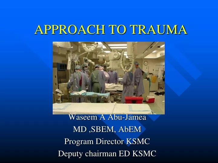
APPROACH TO TRAUMA Waseem A Abu-Jamea MD ,SBEM, AbEM Program Director KSMC Deputy chairman ED KSMC
Objectives • Demonstrate concepts of primary and secondary patient assessment • Establish management priorities in trauma situations • Initiate primary and secondary management as necessary • Arrange appropriate disposition
Trauma • Epidemiology • Leading cause of death in the first 4 decades • 150,000 deaths annually in the US • Permanent disability 3 times the mortality rate • Trauma related dollar costs exceed $400 billion annually
Why ATLS? • Trimodal death distribution • First peak instantly (brain, heart, large vessel injury) • Second peak minutes to hours • Third peak days to weeks (sepsis, MSOF) • ATLS focuses on the second peak…..Deaths from: • TBI, Epidurals, Subdurals, IPH… • Basilar skull fractures, orbital fractures, NEO complex injury… • Penetrating neck injuries… • Spinal cord syndromes… • Cardiac tamponade, tension pneumothorax, massive hemothorax, esophageal injury, diaphragmatic herniation, flail chest, sucking chest wounds, pulmonary contusion, tracheobronchial injuries, penetrating heart injury, aortic arch injuries … • Liver laceration, splenic ruptures, pancreatico-duodenal injuries, retroperitoneal injuries • Bladder rupture, renal contusion, renal laceration, urethral injury… • Pelvic fractures, femur fractures, humerus fractures… • You get the point
Concepts of ATLS • Treat the greatest threat to life first • The lack of a definitive diagnosis should never impede the application of an indicated treatment • A detailed history is not essential to begin the evaluation • “ABCDE” approach
Initial Assessment and Management • An effective trauma system needs the teamwork of EMS, emergency medicine, trauma surgery, and surgery subspecialists • Trauma roles • Trauma captain • Interventionalists • Nurses • Recorder
Primary Survey • Patients are assessed and treatment priorities established based on their injuries, vital signs, and injury mechanisms • ABCDEs of trauma care • A Airway and c-spine protection • B Breathing and ventilation • C Circulation with hemorrhage control • D Disability/Neurologic status • E Exposure/Environmental control
Airway How do we evaluate the airway?
A- Airway • Airway should be assessed for patency • Is the patient able to communicate verbally? • Inspect for any foreign bodies • Examine for stridor, hoarseness, gurgling, pooled secrecretions or blood • Assume c-spine injury in patients with multisystem trauma • C-spine clearance is both clinical and radiographic • C-collar should remain in place until patient can cooperate with clinical exam
Airway Interventions • Supplemental oxygen • Suction • Chin lift/jaw thrust • Oral/nasal airways • Definitive airways • RSI for agitated patients with c-spine immobilization • ETI for comatose patients (GCS<8)
Breathing • What can we look for clinically to assess a patient’s ‘breathing’ status?
B- Breathing • Airway patency alone does not ensure adequate ventilation • Inspect, palpate, and auscultate • Deviated trachea, crepitus, flail chest, sucking chest wound, absence of breath sounds • CXR to evaluate lung fields
Breathing Interventions • Ventilate with 100% oxygen • Needle decompression if tension pneumothorax suspected • Chest tubes for pneumothorax / hemothorax • Occlusive dressing to sucking chest wound • If intubated, evaluate ETT position
What would we do for this patient who is having difficulty breathing?
C- Circulation • Hemorrhagic shock should be assumed in any hypotensive trauma patient • Rapid assessment of hemodynamic status • Level of consciousness • Skin color • Pulses in four extremities • Blood pressure and pulse pressure
Circulation Interventions • Cardiac monitor • Apply pressure to sites of external hemorrhage • Establish IV access • 2 large bore IVs • Central lines if indicated • Cardiac tamponade decompression if indicated • Volume resuscitation • Have blood ready if needed • Level One infusers available • Foley catheter to monitor resuscitation
D- Disability • Abbreviated neurological exam • Level of consciousness • Pupil size and reactivity • Motor function • GCS • Utilized to determine severity of injury • Guide for urgency of head CT and ICP monitoring
Disability Interventions • Spinal cord injury • High dose steroids if within 8 hours • ICP monitor- Neurosurgical consultation • Elevated ICP • Head of bed elevated • Mannitol • Hyperventilation • Emergent decompression
E- Exposure • Complete disrobing of patient • Logroll to inspect back • Rectal temperature • Warm blankets/external warming device to prevent hypothermia
Case • 28 yo M involved in a high speed motorcycle accident. He was not wearing a helmet. He is groaning and utters, “my belly”, “uggghhh”. • HR 134 BP 87/42 RR 32 SaO2 89% on 100% facemask • Brief initial exam: pt is drowsy but arousable to voice, has large hematoma over L parietal scalp, airway is patent, decreased breath sounds over R chest, diffuse abdominal tenderness, obvious deformity to L ankle
ABCDE • What are the management priorities at this time? • What are this patient’s possible injuries? • What are the interventions that need to happen now?
Secondary Survey • AMPLE history • Allergies, medications, PMH, last meal, events • Physical exam from head to toe, including rectal exam • Frequent reassessment of vitals • Diagnostic studies at this time simultaneously • X-rays, lab work, CT orders if indicated • FAST exam
HEENT What are the names of these signs?
Diagnostic Aids • Standard trauma labs • CBC, K, Cr, PTT, Utox, EtOH, ABG • Standard trauma radiographs • CXR, pelvis, lateral C-spine (traditionally) • CT/FAST scans • Pt must be monitored in radiology • Pt should only go to radiology if stable
Tension Pneumothorax How do you treat this?
Hemothorax Is this patient lying or upright?
Widened Mediastinum What disease process does this indicate?
Bilateral Pubic Ramus Fractures and Sacroiliac Joint Disruption What should this injury make you worry about?
Abdominal Trauma • Common source of traumatic injury • Mechanism is important • Bike accident over the handlebars • MVC with steering wheel trauma • High suspicion with tachycardia, hypotension, and abdominal tenderness • Can be asymptomatic early on • FAST exam can be early screening tool
Abdominal Trauma • Look for distension, tenderness, seatbelt marks, penetrating trauma, retroperitoneal ecchymosis • Be suspicious of free fluid without evidence of solid organ injury
Splenic Injury • Most commonly injured organ in blunt trauma • Often associated with other injuries • Left lower rib pain may be indicative • Often can be managed non-operatively Blood from spleen Tracking around liver Spleen with surrounding blood
Liver injury • Second most common solid organ injury • Can be difficult to manage surgically • Often associated with other abdominal injuries Liver contusions
What’s wrong with this picture? • May only see the nasogastric tube appear to be coiled in the lung. • Left > right due to liver protection of the diaphragm. Trace the Diaphragm Outline. Where is the Diaphragm on the left? Abdominal contents Up in the chest on the left
Hollow Viscous Injury • Injury can involve stomach, bowel, or mesentery • Symptoms are a result from a combination of blood loss and peritoneal contamination • Small bowel and colon injuries result most often from penetrating trauma • Deceleration injuries can result in bucket-handle tears of mesentery • Free fluid without solid organ injury is a hollow viscus injury until proven otherwise
bowel mesentery Mesenteric and bowel injury from blunt abdominal trauma. Notice the bowel and mesenteric disruption.
CT Scan in Trauma • Abdominal CT scan visualizes solid organs and vessels well • CT does NOT see hollow viscus, duodenum, diaphram, or omentum well • Some recent surgery literature advocates whole body scans on all trauma • Keep in mind that there is an increase in mortality related to cancer from CT scans
FAST Exam • Focused Abdominal Scanning in Trauma • 4 views: Cardiac, RUQ, LUQ, suprapubic • Goal: evaluate for free fluid See normal Liver and kidney Free fluid in Morrison's Pouch between liver and kidney
momor Morrison’s pouch