Download
1 / 39
420 likes | 659 Vues
Anatomy of Respiratory System. EX 36. Respiration. We can. divide this. into 3 topics. unit. 1) Anatomy. 2). mechanics of breathing. 3). Chemistry of respiration. Respiration. - Consists of 4. events. l). Breathing. (inhalation. exhalation. inspiration /. or. or expiration.

E N D
Respiration We can divide this into 3 topics unit 1) Anatomy 2) mechanics of breathing 3) Chemistry of respiration
Respiration - Consists of 4 events l) Breathing (inhalation exhalation inspiration / or or expiration External respiration 2) exchange of gas between 1 the and blood alveoli the (in the lungs) 3) Internal respiration The exchange of gases from the blood to the tissues cellular respiration 4)
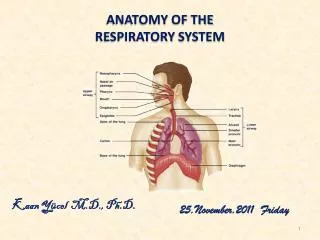
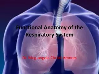
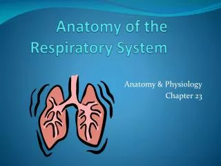
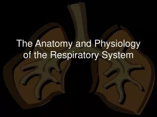
Organization and Functions of the Respiratory System Consists of an upper respiratory tract (nose to larynx) and a lower respiratory tract ( trachea onwards) . Conducting portion transports air. - includes the nose, nasal cavity, pharynx, larynx, trachea, and progressively smaller airways, from the primary bronchi to the terminal bronchioles Respiratory portion carries out gas exchange. - composed of small airways called respiratory bronchioles and alveolar ducts as well as air sacs called alveoli
Respiratory System Functions supplies the body with oxygen and disposes of carbon dioxide filters inspired air produces sound contains receptors for smell rids the body of some excess water and heat helps regulate blood pH
Breathing Breathing (pulmonary ventilation). consists of two cyclic phases: inhalation, also called inspiration - draws gases into the lungs. exhalation, also called expiration - forces gases out of the lungs.
Upper Respiratory Tract Composed of the nose and nasal cavity, paranasal sinuses, pharynx (throat), larynx. All part of the conducting portion of the respiratory system.
Respiratory mucosa A layer of pseudostratified ciliated columnar epithelial cells that secrete mucus Found in nose, sinuses, pharynx, larynx and trachea Mucus can trap contaminants Cilia move mucus up towards mouth
Nose Internal nares - opening to exterior External nares opening to pharynx Nasal conchae - folds in the mucous membrane that increase air turbulence and ensures that most air contacts the mucous membranes
Nose rich supply of capillaries warm the inspired air olfactory mucosa – mucous membranes that contain smell receptors respiratory mucosa – pseudostratified ciliated columnar epithelium containing goblet cells that secrete mucus which traps inhaled particles, lysozyme kills bacteria and lymphocytes and IgA antibodies that protect against bacteria
Nose provides and airway for respiration • moistens and warms entering air • filters and cleans inspired air • resonating chamber for speech detects odors in the air stream rhinoplasty: surgery to change shape of external nose
Pharynx Common space used by both the respiratory and digestive systems. Commonly called the throat. Originates posterior to the nasal and oral cavities and extends inferiorly near the level of the bifurcation of the larynx and esophagus. Common pathway for both air and food.
Pharynx Walls are lined by a mucosa and contain skeletal muscles that are primarily used for swallowing. Flexible lateral walls are distensible in order to force swallowed food into the esophagus. Partitioned into three adjoining regions: nasopharynx oropharynx laryngopharynx
Laryngopharynx Inferior, narrowed region of the pharynx. Extends inferiorly from the hyoid bone to the larynx and esophagus. Terminates at the superior border of the esophagus and the epiglottis of the larynx. Lined with a nonkeratinized stratified squamous epithelium. Permits passage of both food and air.
Lower Respiratory Tract Conducting airways (trachea, bronchi, up to terminal bronchioles). Respiratory portion of the respiratory system (respiratory bronchioles, alveolar ducts, and alveoli).
Larynx Voice box is a short, somewhat cylindrical airway ends in the trachea. Prevents swallowed materials from entering the lower respiratory tract. Conducts air into the lower respiratory tract. Produces sounds. Supported by a framework of nine pieces of cartilage (three individual pieces and three cartilage pairs) that are held in place by ligaments and muscles.
Larynx Nine c-rings of cartilage form the framework of the larynx thyroid cartilage – (1) Adam’s apple, hyaline, anterior attachment of vocal folds, testosterone increases size after puberty cricoid cartilage – (1) ring-shaped, hyaline arytenoid cartilages – (2) hyaline, posterior attachment of vocal folds, hyaline cuneiform cartilages - (2) hyaline corniculate cartlages - (2) hyaline epiglottis – (1) elastic cartilage
Larynx Muscular walls aid in voice production and the swallowing reflex Glottis – the superior opening of the larynx Epiglottis – prevents food and drink from entering airway when swallowing pseudostratified ciliated columnar epithelium
Conduction vs. Respiratory zones Most of the tubing in the lungs makes up conduction zone Consists of nasal cavity to terminal bronchioles The respiratory zone is where gas is exchanged Consists of alveoli, alveolar sacs, alveolar ducts and respiratory bronchioles
Respiratory Bronchioles, Alveolar Ducts, and Alveoli Lungs contain small saccular outpocketings called alveoli. They have a thin wall specialized to promote diffusion of gases between the alveolus and the blood in the pulmonary capillaries. Gas exchange can take place in the respiratory bronchioles and alveolar ducts as well as in the alveoli, each lung contains approximately 300 to 400 million alveoli. The spongy nature of the lung is due to the packing of millions of alveoli together.
Pleura and Pleural Cavities The outer surface of each lung and the adjacent internal thoracic wall are lined by a serous membrane called pleura. The outer surface of each lung is tightly covered by the visceral pleura. while the internal thoracic walls, the lateral surfaces of the mediastinum, and the superior surface of the diaphragm are lined by the parietal pleura. The parietal and visceral pleural layers are continuous at the hilus of each lung.
Pleural Cavities The potential space between the serous membrane layers is a pleural cavity. The pleural membranes produce a thin, serous pleural fluid that circulates in the pleural cavity and acts as a lubricant, ensuring minimal friction during breathing. Pleural effusion – pleuritis with too much fluid
Respiratory events Pulmonary ventilation = exchange of gases between lungs and atmosphere External respiration = exchange of gases between alveoli and pulmonary capillaries Internal respiration = exchange of gases between systemic capillaries and tissue cells
Two phases of pulmonary ventilation Inspiration, or inhalation - a very active process that requires input of energy.The diaphragm, contracts, moving downward and flattening, when stimulated by phrenic nerves. Expiration, or exhalation - a passive process that takes advantage of the recoil properties of elastic fiber. ・The diaphragm relaxes.The elasticity of the lungs and the thoracic cage allows them to return to their normal size and shape.
Muscles that ASSIST with respiration The scalenes help increase thoracic cavity dimensions by elevating the first and second ribs during forced inhalation. The ribs elevate upon contraction of the external intercostals, thereby increasing the transverse dimensions of the thoracic cavity during inhalation. Contraction of the internal intercostals depresses the ribs, but this only occurs during forced exhalation. Normal exhalation requires no active muscular effort.
Muscles that ASSIST with respiration Other accessory muscles assist with respiratory activities. The pectoralis minor, serratus anterior, and sternocleidomastoid help with forced inhalation, while the abdominal muscles(external and internal obliques, transversus abdominis, and rectus abdominis) assist in active exhalation.
LUNG VOLUMES TIDAL VOLUME (TV): Volume inspired or expired with each normalハbreath. = 500 ml INSPIRATORY RESERVE VOLUME (IRV): Maximum volume that can be inspired over the inspiration of a tidal volume/normal breath. Used during exercise/exertion.=3100 ml EXPIRATRY RESERVE VOLUME (ERV): Maximal volume that can be expired after the expiration of a tidal volume/normal breath. = 1200 ml RESIDUAL VOLUME (RV): Volume that remains in the lungs after a maximal expiration.ハCANNOT be measured by spirometry.= 1200 ml
LUNG CAPACITIES INSPIRATORY CAPACITY ( IC): Volume of maximal inspiration:IRV + TV = 3600 ml FUNCTIONAL RESIDUAL CAPACITY (FRC): Volume of gas remaining in lung after normal expiration, cannot be measured by spirometry because it includes residual volume:ERV + RV = 2400 ml VITAL CAPACITY (VC): Volume of maximal inspiration and expiration:IRV + TV + ERV = IC + ERV = 4800 ml TOTAL LUNG CAPACITY (TLC): The volume of the lung after maximal inspiration.ハThe sum of all four lung volumes, cannot be measured by spirometry because it includes residual volume:IRV+ TV + ERV + RV = IC + FRC = 6000 ml