Download
1 / 124
1.24k likes | 1.38k Vues
OA 4.24. Identify the three bones that comprise the shoulder joint. Chapter 16 (pp. 388-407). The Shoulder. introduction. Very complicated region Numerous and varied structures Bones, tendons, muscles, nerves, ligaments, bursae, etc. Highly maneuverable

E N D
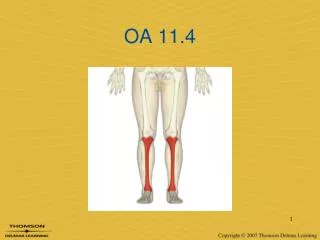
OA 4.24 • Identify the three bones that comprise the shoulder joint.
Chapter 16 (pp. 388-407) The Shoulder
introduction • Very complicated region • Numerous and varied structures • Bones, tendons, muscles, nerves, ligaments, bursae, etc. • Highly maneuverable • 16,000 positions differentiated in 1˚ increments • Combination of four articulations
introduction • Very complicated region • Can be debilitating due to inability to properly use hand(s) • Injuries from: • Direct force/trauma • Secondary forces transmitted proximally • Overuse – predisposition w/overhead movements
introduction • Connection of axial skeleton to upper extremity • Only at SC Joint
stability vs. mobility • The shoulder is inherently unstable • Poor bony stability due to shallow articulation • Stability is gained staticallyby the capsule, ligaments, & labrum; and dynamicallyby the deltoid and rotator cuff muscles.
Stability vs. Mobility CONTINUUM Stabile Mobile As you give up stabilityyou gain mobilityand vice-versa
humerus (proximal) • Landmarks • Head • Greater tubercle (ant. lat.) • Lesser tubercle (ant. med.) • Bicipital groove(between tubercles)
humerus (proximal) • Landmarks • Anatomical neck (proximal to tubes) • Surgical neck (distal to tubes) • Diaphysis • DeltoidTuberosity
scapula • Located between Ribs 2-7 • No direct bony or ligamentous attachments • Articulates with clavicle& humerus • Held against torso by muscleattachment & atmosphericpressure • Functions as both apulley and lever
scapula • Glenoid is oriented about 30-45° anterior to the coronal plane of the body • “Plane of the Scapula” • Places Rotator Cuff muscles at optimal length-tension relationship
scapular landmarks • Body – anterior view = (a) • Subscapular fossa • Medial (vertebral) border • Lateral (axillary) border • Superior Border • Glenoid fossa • Acromion process • Coracoid process
scapular landmarks • Body • Superior angle • Inferior angle
scapular landmarks • Body – posterior view = (b) • Supraspinous fossa • Infraspinous fossa • Greater Scapular Notch • Spine of scapula • Acromion process
scapular landmarks • Body (lateral view) • Glenoid fossa (cavity) • Supraspinous fossa • Acromion process • Coracoid process
scapular landmarks • Body – largest portion of scapula • Rotator cuff attachments - fossa • Separated by borders & angles • Spine of scapula • Separates supraspinous/ infraspinousfossae • Extends to form the acromion process
scapular landmarks • Coracoid process – fingerlike projection towards anterior • Glenoid fossa • Articulates with head of humerus – smaller than head • Superior and inferior tubercles • Surrounded by glenoid labrum
clavicle • “Collarbone” • Slender, S-shaped bone articulating w/ sternum & scapula • Helps maintain alignment of scapula • Anchors upper extremity to axial skeleton
clavicle • Medial 2/3 – more cylindrical • Lateral 1/3 – more flat • Structural Weakness? • Change of direction/shape
articulations • The shoulder is comprised of fourdistinct joints • Sternoclavicular joint • Acromioclavicular joint • Glenohumeral joint • Scapulothoracic joint
sternoclavicular joint (SC) • Articulation of sternum(breastbone) and clavicle at the midline of the body • Anchored by four separate ligaments
acromioclavicular joint (AC) • Articulation of acromion process and clavicle • Three degrees of movement • Elevation/depression • Protraction/retraction • Rotation • Stabilized by three ligaments • Acromioclavicular ligament • Conoid ligament • Trapezoid ligament
glenohumeral joint (GH) • True shoulder joint • Three degrees of freedom • Flexion/extension • ABD/ADD • IR/ER • Combined movements to form • Horizontal flexion/extension • Circumduction • Elevation
glenohumeral joint (GH) • Supported by numerous ligaments that help to form the joint capsule • Superior GH ligament • Middle GH ligament • Inferior GH ligament • Coracohumeral ligament (coracoid process) • Coracoacromial ligament*
GH joint ligaments Conoid Ligament Trapezoid Ligament
GH articular anatomy • Joint capsule allows for ~1cm of movement • >1cm = laxity • The humerus is oriented at the glenoid at a 30o angle of rotation (____version) • < 30o = anteversion • > 30o = retroversion
Scapulothoracic joint (ST) • Not a true joint – no bony articulations • Three degrees of movement • Elevation/depression • Protraction/retraction • Upward/downward rotation • Most important joint for shoulder mechanics • Starting point in the chain of motion • Produces movement at AC & SC joints
scapulohumeral rhythm • The scapula and humerus move at differing degrees during ABD • Humerus moves first 30o • Scapula & humerus move at 1:2 ratio for next 60o • Scapula & humerus move at 1:1 ratio past 90o ABD
ST vs. GH muscles • Scapulothoracic muscles move the scapulaon the axialskeleton • Direct vs. indirect based on attachments • Glenohumeral muscles move the humeruson the scapula
Scapulothoracic muscles • Trapezius • Superior • Middle • Inferior • Levator scapulae • Rhomboids • Major • Minor • Latissimus dorsi
Scapulothoracic muscles • Serratus anterior • Pectoralis major • Pectoralis minor
glenohumeral muscles • Deltoid • Anterior • Middle • Posterior • Triceps brachii • Biceps brachii • Teres major • Coracobrachialis
OA 4.29 • Identify the 4 muscles of the rotator cuff and their corresponding fossae • Identify 3 functions of the rotator cuff
glenohumeral muscles • Rotator cuff muscles • Supraspinatus • Infraspinatus • Teres minor • Subscapularis
rotator cuff muscles • Functions • Holds head of humerus in glenoid fossa • Rotation of humerus on long axis (IR/ER) • Protects shoulder joint via common musculotendinous sheath • Stabilizes shoulder joint in every direction EXCEPTinferiorly
rotator cuff muscles • Rotator Cuff Problems • Rupture of rotator cuff muscles • Calcification of tendons • Humeral subluxation or dislocation • Superiorly: stabilized by supraspinatus & coracoacromial arch • Posteriorly: stabilized by infraspinatus & teres minor • Anteriorly: stabilized by subscapularis • Inferiorly: NO muscular support