Download
1 / 8
80 likes | 105 Vues
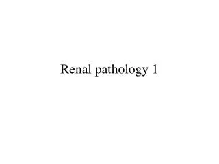
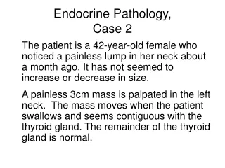
Renal Pathology II, Case 2. A 63-year-old man presents with right flank pain that has been ongoing for the past several months. Identify the organ Describe the gross findings Identify the structures. A – Carcinoma B - Ureter. Renal Cell Carcinoma

E N D
Renal Pathology II,Case 2 • A 63-year-old man presents with right flank pain that has been ongoing for the past several months
Identify the organ Describe the gross findings Identify the structures
A – Carcinoma B - Ureter Renal Cell Carcinoma The surgical specimen consists of a whole kidney and attached ureter. There is a large, irregular carcinoma in the superior pole of the kidney.
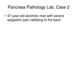
Identify the organ Describe the gross findings Identify the structures
A – Carcinoma B – Normal Cortex Renal Cell Carcinoma The surgical specimen consists of a bisected kidney that contains a renal cell carcinoma in the superior pole. The large, irregular neoplasm is yellow to golden and contains areas of hemorrhage (red). The yellow color and hemorrhage (increased vascularity) are characteristic pathologic findings in a renal cell carcinoma. Correlate the pathologic change with possible clinical presentations.
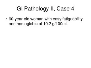
Describe the gross findings Identify the structures. Diagnosis?
A – Carcinoma B – Normal cortex C – Ureter D - Aorta Renal Cell Carcinoma The autopsy specimen consists of a bisected left kidney, normal right kidney (arrow) and attached aorta. A renal cell carcinoma occupies the superior pole of the left kidney. The neoplasm is yellow and contains extensive hemorrhage
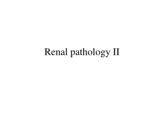
Describe the low and high power histologic findings of sections from the mass Renal cell carcinoma, clear cell type Tumor cells are rounded or polygonal shaped with abundant clear or granular cytoplasm (on special stains the cytoplasm stains for fat and glycogen).