Download
1 / 16
170 likes | 241 Vues
Case Study 63: Cancer of the Female Breast. By Robyn Schwartz. Case Background. 46, premenopausal Dense breasts Has noticed cysts in the past Noticed new lump in upper right quadrant Did not resolve Got bigger Denied lumps in axillary. Patient history. Happily married for 21 years

E N D
Case Study 63: Cancer of the Female Breast By Robyn Schwartz
Case Background • 46, premenopausal • Dense breasts • Has noticed cysts in the past • Noticed new lump in upper right quadrant • Did not resolve • Got bigger • Denied lumps in axillary
Patient history • Happily married for 21 years • 3 kids (3, 8, and 10) • Does breast self exams • Normal pap 2 years ago • Has Asthma and hypertension • Exercises • No tobacco, alcohol or illegal drugs
Risk Factors • All 3 kids born after the age of 35 • First period at 11yr old • Dense breasts • Cysts already develop regularly • Family history of breast cancer • Paternal grandmother diagnosed at age 45 before menopause • Mother diagnosed at age 45 before menopause. Died at age 73 from reoccurrence of breast cancer
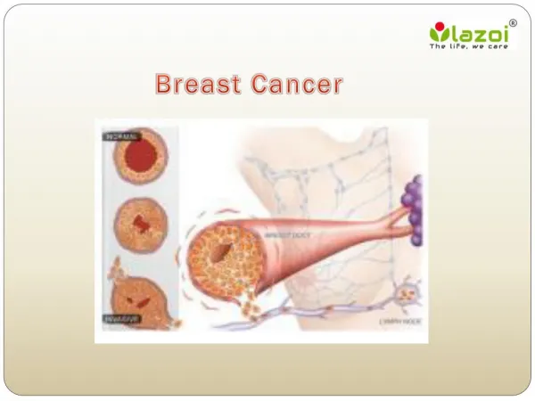
Breast Cancer: What is it? • Uncontrolled division of abnormal cells in the breast • Caused by specific mutations • BRCA1 and BRCA2 • TP53
Our patient: Mammogram • 2.3cm x 2.9cm x 3.2cm mass • Irregular borders • Skin thickening • Enlarged axillary lymph node • 6 Y-shaped microcalcifications extended toward nipple • Abnormal mass into pectoral muscle
Grading vs Staging • How far the cancer has spread • I, II, III, IV • Based on • Size of tumor • Invasive vs non invasive • Spread to lymph nodes • Spread to other parts of the body • How abnormal the cells are • 1, 2, 3,4 • Based on • Tubule formation • Size and shape of cells • Mitotic division • Measures the likely aggressiveness of the cells
Grading tumors Nuclear (size/shape) Score 1: little variation in size Score 2: moderate variability, open vesicular nuclei Score 3: lots of variability open nuclei Mitotic Score 1: <7 mitoses Score 2: 8-14 mitoses Score 3: >14 mitoses Tubular differentiation Score 1: > 75% glandular/tubular Score 2: 10-75% glandular/tubular Score 3: < 10% Glandular/ tubular
Staging Cancer • agrdjdytydstasf Stage 0: No Cancer Stage I: IA: Cancer is small, low grade and localized IB: Cancer is large, low grade and localized Stage II: IIA: Tumor is 2-5cm but has not spread IIB: Tumor is 2-5cm but has spread to lymph nodes Stage III: Tumor is larger than 5cm and has spread to multiple lymph nodes Stage IV: Cancer has spread to other parts of the body
Our Patient: Biopsy and ultrasound • Ultrasound: • Non-cystic mass, solid appearing • Abnormal vascularity • Some skin thickening and mild tissue edema • Biopsy: • Consistent with infiltrating breast cancer • 3-5 divisions per high power field • Mild pleomorphism • Positive for estrogen and progesterone receptors
Grade and Stage • Grade 1 • Mitotic score: 1 (<7 divisions) • Glandular Score: 1 (75% glandular) • Nucleic Score: 1 (not much change) • Total score: 3 • 10 year survival rate 90% • Stage IIB • Small • Spread to 1 lymph node • 5 year survival rate of 71%
Our Patient: Treatment • Breast conservation therapy • Lump removal • Radiation • Lymph node biopsy • Tamoxifen • Estrogen receptor blocker • Helps stop growth of cancer cells
Our Patient: Follow Up • 6.5 years cancer free • 80 months later, complained of • bone pain in lower back • Headache
Test Results • Bone scan • Lesions in lumbar spine without fracture • Chest X-Ray • 3 small nodules in upper lobe of left lung • Brain MRI • Small mass in right frontal lobe • Abdominal CT • Negative • Blood tests • CEA elevated by 2-fold • CA27-29 concentration elevated by 2-fold
Diagnosis, Outlook, and Treatment • Stage IV Breast cancer • 13% 10-year survival rate • Treatments • Chemotherapy • taxanes • Hormone Therapy • Targeted therapy • HER2 targeted therapy • Slow growth • Manage Bone Metastasis • Biophosphonates • Slow destruction
How to Prevent cancer • Exercise • Eat well • Don’t smoke • Do regular breast self exams • Report anything suspicious immediately • Check family history