Download
1 / 38
380 likes | 391 Vues
Structure and Characteristics of Muscle. 1.https://www.youtube.com/watch?v=ousflrOzQHc https://www.youtube.com/watch?v=qLDC6-PH6rA 2. https://www.youtube.com/watch?v=sIH8uOg8ddw 3. https://www.youtube.com/watch?v=FfFBoIkdDgQ. Introduction.

E N D
Structure and Characteristics of Muscle 1.https://www.youtube.com/watch?v=ousflrOzQHc https://www.youtube.com/watch?v=qLDC6-PH6rA 2. https://www.youtube.com/watch?v=sIH8uOg8ddw 3. https://www.youtube.com/watch?v=FfFBoIkdDgQ
Introduction • Tendon is fibrous connective tissue (non elastic) which is thick with while colour connecting skeletal muscle with bones. • Ligament is fibrous connective tissue connecting bone with other bone. • Muscle is skeletal muscle fibre which is a bundle of fasciculus (cylindrical-shaped muscle bundles bound by connective tissue.
Differences between Tendon and Ligament • Both are connective tissue;tendon connects muscle with bone, while ligament connects bone with bone. • Tendon connects the edge of muscle for bone, while ligament always connects bones in their joints. • There is only one tendon for certain muscle in one edge, while there are several ligaments connecting two bones in one joint. • Tendon has more collagen compared to ligament. • Ligament has more proteoglycancompared to tendon. • Tendon has higher blood supply compared to ligament.
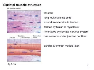
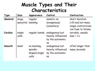
Types of muscle tissue • Skeletal muscle: striated muscle fibre, has multiple nuclei, innervated by somatic motor neurons (voluntary), and attached to bone • Smooth muscle: smooth muscle fibre (non striated), has one nucleus located in the middle, innervated by autonomic nervous (involuntary), found in the internal organ (viscera) • Cardiac muscle: smooth muscle which is involuntary; striated muscle, has one nucleus, innervated by autonomic nervous (involuntary)
Structure of Muscle • Muscle • Muscle bundle/ fascicle • Muscle fiber/ cells • Fibril (myofibril) • Filament (myofilament) • Thick filament (myosin) and thin filament (actin).
Connective tissue on meat • Epimysium : connective tissue that surrounds muscles • Perimysium : connective tissue that surrounds muscle fiber bundle • Endomysium: connective tissue that surrounds muscle fiber.
Muscle Fiber • Structural unit of skeletal muscle called as muscle fiber/ muscle cell. • Muscle fiber composes 75-92% of muscle, the remaining consists of connective tissue, blood vessels, nervous tissues, and extracellular fluid. • Muscle fiber in mammals and avian is long, unbranched, and multinucleated. • Muscle fiber has a diameter of 10-100m.
Sarcolemma • Sarcolemma is membrane covering muscle fiber. • Sarcolemma forms tubule called transverse tubules or T system. • Sarcolemma is elastic, and functions in muscle contraction and relaxation.
Sarcoplasm • Sarcoplasm is the cytoplasm of muscle fiber. • It is intracellular colloid substance composed of water as much as 75-80%, and the remaining consists of lipid, glycogen granules , ribosome , protein , NPN and inorganic components.
Nuclei • Skeletal muscle fiber has multiple nuclei, because the shape of the cell is long. • Nuclei can be found every 5 m of distance.
Myofibril • Myofibril is organelle of cylindrical, long, and thin muscle fiber with a diameter of 1-2 m. • The length of myofibril is parallel to the length of muscle fiber. • Muscle fiber whose diameter is 50 m contains 1000-2000 myofibril. • Myofibril consists of segments called sarcomere. • The length of sarcomere is approximately 2,5 m.
Myofilament • Sarcomere contains thick filament (diameter of 14-16 nm and length of 1,5 m) and thin filament (diameter of 6-8 nm and length of 1 m). • Thick filament forms A-band and consist of myosin protein. • Thin filament consists of I-band and consists of actin protein .
Myofibril Protein • It consists of more than 20 types of myofibril protein. • There 6 main proteins: myosin, actin, titin, tropomyosin, troponin and nebulin. • The functions of myofibril protein are categorized as contractile proteins , regulatory proteins and cytoskeletal protein.
Myofibril Protein • Myofibril protein functioning as the main contractile protein is actin and myosin. • Myofibril protein functioning as the main regulatory protein is tropomyosin and troponin. • Myofibril protein functioning as cytoskeletal protein is titin and nebulin.
Actin • Actin protein composes of approximately 20% of the total myofibril protein. • Actin is round globular protein which is also called as G-actin with a diameter of 5,5 nm. • Fibrous actin is formed when G-actin undergoes polymerization forming actin chain. • Two actin chains form spiral then form super helix (actin filament).
Myosin • Myosin composes approximately 45% from the total myofibril protein. • Myosin is fibrous protein. • Myosin is thin and long, and a little thick on its edge. • The thick part of the edge is called head, the long part is (backbone) is tail, and the part within head and tail is called neck. • Myosin undergoing hydrolysis will result in 2 main fractions, namelymyosin heavy chain and myosin light chain..
Tropomyosin • Tropomyosin composes approximately 5% from the total myofibril protein. • Tropomyosin is located near actin filament. • One molecule of tropomyosin is found along 7 molecule of G-actin in actin filament.
SarcoplasmicProtein • Enzymes related to glycolysis process (73%). • Enzymes related to creatin kinase (9%). • Myoglobin (meat pigment). • Hemoglobin (blood pigment).
Myoglobin • Myoglobin is a single proteinchain with 153 amino acid residues with heme group which can store oxygen. • Myoglobin has higher affinity for oxygen compared to haemoglobin. • The existence of myoglobin in meat can result in red color of meat. • Heme group binds to nitrogen from histidine and iron from that heme.
Hemoglobin • Hemogobin has a structure which is almost similar to that of myoglobin, • Hemoglobin has quarternarystructure because it consists of 4 sub-units of protein chain. • Each sub-unit of protein chain has heme group so that each molecule of hemoglobin contains 4 molecules of oxygen. • Reacting with oxygen to form oxyhemoglobin • Oxygenation of hemoglobin occurs in blood when blood passes lungs.
Protein of Connective Tissue • Distributed to all components of body: muscle, blood and lymph vessels, tendon, nerves, and skin. • Withstanding infectious agents. • Protecting fat-storing cells (adipose tissue). • Protecting muscles, muscle fiber bundle, and muscle fiber.
Protein of Connective Tissue • Connective tissue is composed of ground substance, cells, and extracellular fibers. • Ground substance is viscous solution containing soluble glycoprotein, substrate, and end product of connective tissue metabolism (tropocollagen and tropoelastin). • The cells of connective tissue consist of fixed cells and wandering cells. • Extracellular fiber is composed of collagen, elastin and reticulin.
Collagen • Collagen composes approximately 20-25% from the total protein in the body. • Collagen is structural protein in connective tissue. • Collagen has direct effect on meat tenderness. • The amount and strength of collagen increase with increasing age.
Collagen • Collagen is the main component of skin tissue, tendon, ligament, bone and cartilage. • Collagen fiber has a diameter of 1-12 m. • Collagen contains amino acid specific hydroxyprolin in a relatively fixed amount, so that the level of hydroxyprolin can be used to determine the amount of collagen tissue.
Collagen • Collagen fibril is formed from tropocollagen molecules. • Tropocollagen is protein with BM 300 kDa, and is formed of 3 polypeptide chains that overlap to form triplehelix. • The process of collagen formation requires vitamin C, i.e. during the hydroxylation of prolin into hydroxyprolin.
Muscle Contraction • Muscle contraction is the process of muscle shrinkage/Shortening that needs energy. • The proteins involved in the contraction process are actin, myosin, tropomyosin dan troponin. • The energy for this process is from ATP. • The process of muscle contraction consists of contraction (muscle shortening) and relaxation (muscle stretching)
Stages of Contraction Process • Stimulus from nerve to neuromuscular junction ending. • Action potential causes acetylcholine to be released to the surface of the muscle fibers. • Action potential spreads to all muscle fibers through T-system (transverse tubule). • Action potential is sent to SR and SR releases Ca2+ ion.
Stages of Contraction Process • Ion Ca2+ activates troponin and tropomyosin in thin filament, forming a strand between actin and myosin; filament shift occurs which then leads to muscle contraction
Stages of Relaxation Process • It starts with partial repolarization of action potential. • Acetylcholine at the neuromuscular junction is broken down by acetylcholinesterase, causing the termination of the stream of action potentials along the muscle fiber surface • SR ceases to release Ca2+ ions used for contraction.
Stages of Relaxation Process • Troponin releases Ca2+ ions so that change in the configuration of troponin and tropomyosin occurs. then blocks the action of the myosin molecule heads, and contraction ceases. • The activity of molecule heads of myosin and the formation of strand between actin and myosin are blocked so that contraction ceases.