Download
1 / 36
400 likes | 756 Vues
Forensic Anthropology. Forensic Anthropology William M. Bass, Ph. D. Professor Emeritus Anthropology Department University of Tennessee. Forensic Anthropology. Dr. T. Dale Stewart “Essentials of Forensic Anthropology” (1975)

E N D
Forensic Anthropology Forensic Anthropology William M. Bass, Ph. D. Professor Emeritus Anthropology Department University of Tennessee
Forensic Anthropology Dr. T. Dale Stewart “Essentials of Forensic Anthropology” (1975) “that branch of physical anthropology which, for forensic purposes, deals with the identification of more or less skeletonized remains known to be, or suspected of being human.”
Coroners/Medical Examiners/Forensic Pathologists • To investigate unexplained civilian deaths, • Are trained primarily to deal with “fresh” remains
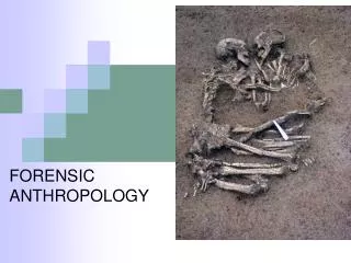
Forensic Anthropologist • Usually last person in the human identification chain • Most bodies can be identified by morphological features or fingerprints • Bodies burned or decomposed beyond recognition require the expertise of a trained osteologist
Anthropology • That field of science that studies man from his earliest beginnings on the earth (about 5 million years ago) to the present • The scientific study of the skeleton (both human and animal) • The application of these skills to medico-legal cases if forensic anthropology
The Big Four • Determining age at death • Determination of sex • Determination of race • Determination of stature • A basic knowledge of human biology, growth and development, and the aging process is needed to identify gradual changes that occur in the skeleton from birth to old age.
Determination of Age At Death • Aging divided into two major categories • Maturation and Degeneration
I. Maturation • Dentition—man is a mammal and has 2 sets of teeth • Deciduous • Adult
Deciduous (Baby or milk teeth) • Approx 6 mo of age: lower central incisors erupt • Approx 24 mo of age: all 20 deciduous teeth are usually erupted • Approx 2-6 yrs: retain deciduous teeth, but as skeleton grows, children develop spaces between teeth (teeth don’t get larger, but bone does grow)
Adult Teeth • Larger in size and whiter in color (thinner enamel) • Approx 6 yrs: first adult tooth, six year molars erupt behind the 20 deciduous teeth • Approx 6 ½ -11 ½ years: baby teeth replaced by adult teeth • Approx 12 yrs: 2nd molars erupt • Approx 18 yrs: 3rd molars may or may not erupt; genetically we are losing 3rd molars; many are impacted or never erupt; any number of 3rd molars may be missing
I. Maturation • Epiphyses • During growth process there are 806 centers of ossification which unite into 206 bones • By age 13 in girls and 15 in boys, the epiphyses begin to unite to the diaphyses (shaft) • From 13-17 yrs most active period of epiphyseal union; major growth of body stops with closure of epiphyses • Age 25: last epiphysis to unite is the sternal end of the clavicle • During growth process, age can be determined by measuring length of any long bone and comparing with published standards (Trotter and Gleser 1958)
II. Degenerative Changes • Following end of growth period the skeleton begins a slow process of wearing out • Degenerative changes appear in early 20s and present in increasing degrees until death • 3 best areas to estimate age at death of an adult are: • Pubic Symphysis • Osteon Counting • Osteoarthritic Lipping
A. Changes in Pubic Symphysis • Essentially what happens is that as one grows older, the face of the pubic symphysis changes from one that is “rough” with an appearance of “mountains” and “valleys” to one that becomes smoother with the “mountains” wearing off and the “valleys” filling up. • One caution—late in life changes of the pubic symphysis becomes more obscure and determination of age at death in the 60’s and 70’s with a +/- 5 year range is difficult.
B. Osteon Counting • Microscopic method developed by Ellis R. Kerley in the 1950’s • Requires sectioning long bone at its midshaft, cutting, grinding, and polishing a thin section (80 microns) and viewing this section through polarized light.
C. OsteoarthriticLipping • As individuals age, their skeletons reflect the various stresses encountered throughout life
Determination of Sex • Skeletal sexual criteria manifest themselves at puberty and are not clear in the skeleton below age of 12-1 5 • Pelvis • Skull • Rest of the skeleton
Pelvis • Women have broader hips than men • Width attributed to 3 areas: • Length of pubic bone: female has long pubic portion and a much wider subpubic angle • Width of sciatic notch: notch is narrow in men and wide in females • Bone build-up on the sacroiliac joint: portion of the ilium where it articulates with the sacrum is flat in males; there is a build-up of bone in this area in a female
Skull • Sex differences in the skull are mainly due to sexual dimorphism where the male is larger, more rugged and muscle marked; whereas, the female is smaller, more gracile, and smooth • Males have larger mastoid processes, well-marked supra-orbital ridges, prominent muscle markings in the occipital region, and metrically a larger mouth • Females usually have a smooth forehead with little or no supra-orbital ridges with shallow muscle markings on the occipital • The male chin is usually square, while it comes to a point in a female
The Rest of the Skeleton • Males are usually larger in size because they normally reach puberty 2 years later than females and thus grow 2 years longer • Overall size is the main criteria but specific areas that should be looked at are: • Maximum Diameter of the Head of the Femur • Width of the Ala of the Sacrum • Length of the Sternum • Septal Aperatures of the Humerus
The Rest of the Skeleton • Maximum Diameter of the Head of the Femur—a measurement below 43mm suggests female, above 45mm a male • Width of the Ala of the Sacrum—width of the articular area of the sacrum in a male will be greater than half the width of the entire sacrum and in females is less than half the width of the sacrum • Length of the Sternum—combined length of 121mm or less included only females, and sternal lengths of 173mm and above included only males • Female x-142mm • ? 143-157mm • Male 158-xmm • SeptalAperatures of the Humerus—presence of a supra condyloid foramen in the distal end of the humerus has long been known to have a greater frequency in females
Determination of Race • Anthropometric Measurements • In 1962, Eugene Giles and Orville Elliot published information on the use of anthropometric measurement to determine race of a skull. This procedure allows a discrimination between Caucasoids (Whites), Negroids (Blacks), and Mongoloids (American Indians).
Determination of Race • Prognathism (in Negroid skulls) • Prognathism is protrusion of the alveolar regions of the mandible and maxilla. It is common in Negroid (Black) individuals, is seen slightly in Mongoloids, and only occasionally in Caucasoids (Whites). • A quick test….
Determination of Race • Nasal Sill (in Caucasoid skulls) • Observe the base of the nasal apature • A quick test….
Determination of Race • Flat Face (in Mongoloid skulls) • Usually Mongoloids, (including American Indians), they have a flat, usually round face. Faces of Whites come to a point along the midline. • A quick test….
Determination of Race • Edge to Edge Bite in Incisor Region • Occlude your teeth, most will have an overbite, a situation where the upper or maxillary incisors are in front of the lower or mandibular incisors. • Mongoloids, and especially American Indians, have an edge to edge bite in the incisor region. • If you have a skull with the incisor teeth present, look for wear on the occlusal (biting) surface of the incisor teeth. If wear is present i.e., the enamel is worn off, then you have an American Indian skull.
Determination of Stature • A number of formulae in exist which enable calculation of stature from long bones. • In general, most calculations are based on the maximum length of the long bones. • Probably the best method of use on skeletal material from the US are formulae developed by Trotter and Gleser (1958) on Whites, Blacks, Mongoloids, and Mexicans. • Measurement of long bones must be in centimeters.