Download
1 / 7
70 likes | 242 Vues
The effect of angular extent on reconstructed image quality in tomosynthesis. A tissue sample example. Tissue Imaging Example. Lumpectomy, embedded in agar 47F, node negative 3.5 cm dia.; IDC, grade III. 67 mm. Whole Mount Histology Slide. Lumpectomy. Constant Exposure/Projection.

E N D
The effect of angular extent on reconstructed image quality in tomosynthesis A tissue sample example
Tissue Imaging Example • Lumpectomy, embedded in agar • 47F, node negative • 3.5 cm dia.; IDC, grade III 67 mm Whole Mount Histology Slide
Lumpectomy Constant Exposure/Projection CT (P=93, 186°)300μm× 300 μm × 600 μm voxelsRelative Dose: 10 Tomosynthesis (P=11, 22°)300μm× 300 μm × 600 μm voxelsRelative Dose: 1
Tissue Imaging Angular Spacing, Δθ=2°
More artifacts, Background tissue partly suppressed Less clutter in background, better contrast
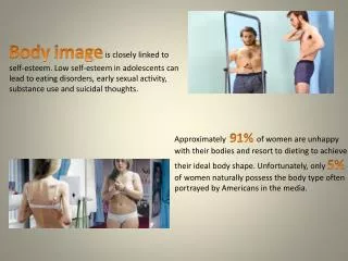
Background complexity phantom 6 acetate beads (1.5-8 mm) Natural sea sponge (including coral pieces) in corn oil Phantom Development Evaluating the Effect of Dose on Reconstructed Image Quality in Digital Breast Tomosynthesis, M. P. Kempston et al., IWDM 2006 Poster Session