Download

1 / 28
320 likes | 1.06k Vues
SUBCLAVIAN ARTERY AND INTERNAL JUGULAR VEIN. Miss.D.Blessina Sugandhi. INTRODUCTION. It is the principal artery which continues as axillary artery for the upper limb. It also supplies a considerable part of the neck and brain through its branches. ORIGIN. On the right side,

E N D
SUBCLAVIAN ARTERY AND INTERNAL JUGULAR VEIN Miss.D.Blessina Sugandhi
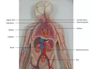
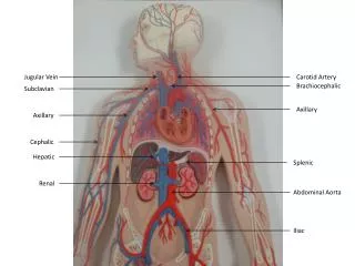
INTRODUCTION • It is the principal artery which continues as axillary artery for the upper limb. • It also supplies a considerable part of the neck and brain through its branches.
ORIGIN • On the right side, It is branch of bracheocephalic artery • On the left side, it is branch of arch of aorta • Both arteries enter the neck by passing behind the sternoclavicular joints • And they pursue similar course in the neck
COURSE • Each artery arches laterally from the sternoclavicular joint rib where it ends by becoming continuous with the axillary artery • The scalenus anterior muscle crosses the artery anteriorly and divides it into 3 parts • The first part is medial, the second part posterior, and the third part lateral to the scalenus anterior muscle
RELATIONS 1ST PART Anteriorly – (Medial to lateral) - Common carotid artery - Vagus nerve - Internal jugular vein - Sternothyoid and sternohyoid muscles - Sternocleidomastoid muscle Posteriorly – - Suprapleural membrane - Cervical pleura - Apex of lung
2nd part Anteriorly - Scalenus anterior - Right phrenic nerve - Sternocleidomastoid muscle Posteriorly - Suprapleural membrane - Cervical pleura - Apex of lung
Superiorly - Upper and middle trunks of brachial plexus Inferiorly - First rib 3rd Part Anteriorly - Middle 1/3rd of clavicle - Post. Border of sternocleidomastoid muscle Posteriorly - Scalenus medius - Lower trunk of brachial plexus - Suprapleural membrane - Cervical pleura - Apex of lung
BRANCHES 1ST PART - 1. Vertebral artery 2. Internal thoracic artery 3. Thyrocervical trunk 4. Costocervical trunk (Left side) 2ND PART - 1. Costocervical trunk (Right side) 3RD PART - Dorsal scapular artery (Occasionally)
Costocervical Trunk Right side – Post. Surface of 2nd part of subclavian artery Left side – First part of the artery
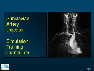
APPLIED ASPECT • The 3rd part of the subclavian artery can be effectively compressed against the 1st rib after depressing the shoulder • A cervical rib may compress the subclavian artery, diminishing the radial pulse
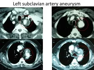
The right subclavian artery may arise from the descending thoracic aorta. In that case, it passes posterior to the oesophagus which may be compressed and the condition is known as dysphagialusoria • An aneurysm may form in the 3rd part of the subclavian artery. Its pressure on the brachial plexus causes pain, weakness, and numbness in the upperlimb
Subclavian steal syndrome Obstruction to the subclavian artery proximal to the origin of vertebral artery may lead to stealing of blood from the brain through the opposite vertebral artery. This may provide necessary blood to the affected side. The nervous symptoms incurred are called “subclavian steal syndrome”.
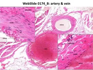
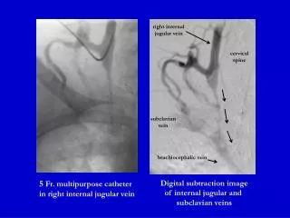
Internal Jugular Vein • ORIGIN AND COURSE • Collects most venous blood from head and neck • Begins in jugular foramen, as a continuation of sigmoid sinus ends posterior to corresponding sternoclavicular joint, where it joins subclavian vein to form brachiocephalic vein • Lies within carotid sheath, lateral to internal carotid artery (in upper neck), and lateral to common carotid artery (in lower neck) • Deep cervical lymph nodes lie alongside internal jugular vein • Upper and lower ends of internal jugular vein are dilated (superior and inferior bulbs)
TRIBUTARIES: • inferior petrosal sinus leaves cranial cavity via jugular foramen and drains into superior bulb • lingual vein(s) • pharyngeal veins • facial vein • superior and middle thyroid veins • occipital vein (sometimes)
Anteriorly - Stenocleidomastoid - Posterior belly of digastric muscle - Superior belly of omohyoid muscle - Parotid gland - Styloid process Posteriorly - Transverse process of atlas - Cervical plexus - Scalenus anterior muscle - First part of subclavian artery Medially - Internal carotid artery - Common carotid artery - Vagus nerve
The thoracic duct opens into the angle of union between the left internal jugular vein and the left subclavian vein. The right lymphatic duct opens similarly on the right side. In the middle of the neck, the internal jugular vein may communicate with theexternal jugular vein through the oblique jugular vein which runs across the anterior border of the sternocleidomastoid.
CLINICAL ASPECTS • Deep to the lesser supraclavicular fossa, the internal jugular vein is easily accessible for recording of venous pulse tracings. The vein can be cannulated by direct puncture in the interval between the sternal and clavicular heads of sternocleidomastoid muscle • In congestive cardiac failure or any other disease where venous pressure is raised, the internal jugular vein is markedly dilated and engorged.