Download
1 / 28
280 likes | 862 Vues
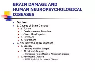
Animal Models of Human Neuropsychological Diseases. Experiments regarding neuropathology are not usually possible with human subjects Animal models are often utilized, for example: Kindling model of epilepsy Experimentally induced seizure activity Transgenic mouse model of Alzheimer’s

E N D
Animal Models of Human Neuropsychological Diseases • Experiments regarding neuropathology are not usually possible with human subjects • Animal models are often utilized, for example: • Kindling model of epilepsy • Experimentally induced seizure activity • Transgenic mouse model of Alzheimer’s • Mice producing human amyloid • MPTP model of Parkinson’s • Drug-induced damage comparable to that seen in PD
Kindling Model of Epilepsy • A series of periodic brain stimulations eventually elicits convulsions – the kindling phenomenon • Neural changes are permanent • Produced by stimulation distributed over time • Convulsions are similar to those seen in some forms of human epilepsy – but they only occur spontaneously if kindled for a very long time • Kindling phenomenon is comparable to the development of epilepsy (epileptogenesis) seen following a head injury
Transgenic Mouse Model of Alzheimer’s Disease • Only humans and a few related primates develop amyloid plaques • Transgenic – genes of another species have been introduced • Genes accelerating human amyloid synthesis introduced into mice • Plaque distribution comparable to that in AD • Unlike humans, no neurofibrillary tangles
MPTP Model of Parkinson’s Disease • The Case of the Frozen Addicts • Synthetic heroin produced the symptoms of Parkinson’s • Contained MPTP • MPTP causes cell loss in the substantia nigra, like that seen in PD • Animal studies led to the finding that deprenyl can retard the progression of PD
Neuroplastic Responses to Nervous System Damage • Degeneration – deterioration • Regeneration – regrowth of damaged neurons • Reorganization • Recovery
Degeneration • Cutting axons (axotomy) is a common way to study responses to neuronal damage • Anterograde: degeneration of the distal segment – between the cut and synaptic terminals • Cut off from cell’s metabolic center – swells and breaks off within a few days • Retrograde: degeneration of the proximal segment – between the cut and cell body • Progresses slowly – if regenerating axon makes a new synaptic contact, the neuron may survive
FIGURE 10.15 Neuronal and transneuronal degeneration following axotomy.
Neural Regeneration • Does not proceed successfully in mammals and other higher vertebrates – capacity for accurate axonal growth is lost in maturity • Regeneration is virtually nonexistent in the CNS of adult mammals and unlikely, but possible, in the PNS
Neural Regeneration in the PNS • If the original Schwann cell myelin sheath is intact, regenerating axons may grow through them to their original targets • If the nerve is severed and the ends are separated, they may grow into incorrect sheaths • If ends are widely separated, no meaningful regeneration will occur
FIGURE 10.16 Three patterns of axonal regeneration in mammalian peripheral nerves.
Mammal PNS Neurons Regenerate, CNS Don’t • CNS neurons can regenerate if transplanted into the PNS, while PNS neurons won’t regenerate in the CNS • Schwann cells promote regeneration • Neurotrophic factors stimulate growth • CAMs provide a pathway • Oligodendroglia actively inhibit regeneration
Collateral Sprouting When an axon degenerates, axon branches grow out from adjacent healthy neurons & synapse at vacated sites
Neural Reorganization • Reorganization of primary sensory and motor systems has been observed in laboratory animals following • Damage to peripheral nerves • Damage to primary cortical areas • Lesion one retina and remove the other – V1 neurons that originally responded to lesioned area now responded to an adjacent area – remapping occurred within minutes • Studies show large scale of reorganization possible
Cortical Reorganization Following Damage in Humans • Brain-imaging studies indicate there is continuous competition for cortical space by functional circuits • e.g. Auditory and somatosensory input may be processed in formerly visual areas in blinded individuals
Mechanisms of Neural Reorganization • Strengthened existing connections due to a release from inhibition? • Consistent with speed and localized nature of reorganization • Establishment of new connections? • Magnitude can be too great to be explained by changes in existing connections
2-Stage Model of Neural Reorganization Before damage 1. Strengthening of existing connections thru release from inhibition 2. Establishment of new connections by collateral sprouting.
Recovery of Function after Brain Damage • Difficult to conduct controlled experiments on populations of brain-damaged patients • Can’t distinguish between true recovery and compensatory changes • Cognitive reserve – education and intelligence – thought to play an important role in recovery of function – may permit cognitive tasks to be accomplished in new ways • Adult neurogenesis may play a role in recovery
FIGURE 10.21 Increased neurogenesis in the dentate gyrus following damage (These images are courtesy of Carl Ernst and Brian Christie, Department of Psychology, University of British Columbia.)
Neuroplasticity and the Treatment of Nervous System Damage • Reducing brain damage by blocking neurodegeneration • Promoting recovery by promoting regeneration • Promoting recovery by transplantation • Promoting recovery by rehabilitative training
Reducing Brain Damage by Blocking Neurodegeneration • Various neurochemicals can block or limit neurodegeneration • Apoptosis inhibitor protein – introduced in rats via a virus • Nerve growth factor – blocks degeneration of damaged neurons • Estrogens – limit or delay neuron death • Neuroprotective molecules tend to also promote regeneration
Promoting CNS Recovery by Promoting Regeneration • While regeneration does not normally occur in the CNS, experimentally it can be induced directing growth of axons by • Schwann cells • Olfactory ensheathing cells
Promoting Recovery by Neurotransplantation • Transplanting fetal tissue • Fetal substantia nigra cells used to treat MPTP-treated monkeys (PD model) • Treatment was successful • Limited success with humans • Transplanting stem cells • e.g. Embryonic stems cells implanted into damaged rat spinal cord • Rats with spinal damage with improved mobility
Promoting Recovery by Rehabilitative Training • Monkeys recovered hand function from induced strokes following rehab training • Constraint-induced therapy in stroke patients – tie down functioning limb while training the impaired one – creates a competitive situation to foster recovery • Facilitated walking as an approach to treating spinal injury
Promoting Recovery by Rehabilitative Training Continued • Benefits of cognitive and physical exercise • Correlations in human studies between physical/cognitive activity and resistance or recovery from neurological injury and disease • Rodents raised in enriched environments are resistant to induced neurological conditions (epilepsy, models of Alzheimer’s, Huntington’s, etc.) • Physical activity promotes adult neurogenesis in rodent hippocampus
Phantom Limbs: Neuroplastic Phenomena • Ramachandran’s hypothesis: phantom limb caused by reorganization of the somato-sensory cortex following amputation • Amputee feels a touch on his face and also on his phantom limb (due to their proximity on somatosensory cortex) • Amputee with chronic phantom limb pain gets relief through visual feedback: view in mirror of his intact hand unclenching as seen in mirror box
FIGURE 10.23 The places on Tom’s body where touches elicited sensations in his phantom hand. (Based on Ramachandran & Blakeslee, 1998.)